1M4H

Crystal Structure of Beta-secretase complexed with Inhibitor OM00-3


Experimental Data Snapshot

  • Method: X-RAY DIFFRACTION
  • Resolution: 2.10 Å
  • R-Value Free: 0.271 
  • R-Value Work: 0.216 

wwPDB Validation   3D Report Full Report


This is version 2.0 of the entry. See complete history


Literature

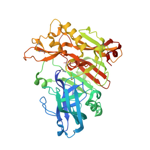
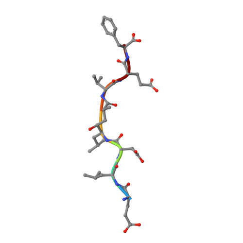
Crystal Structure of Memapsin 2 (beta-Secretase) in Complex with Inhibitor OM00-3

Hong, L.Turner, R.T.Koelsch, G.Shin, D.Ghosh, A.K.Tang, J.

(2002) Biochemistry 41: 10963-10967

  • DOI: https://doi.org/10.1021/bi026232n
  • Primary Citation of Related Structures:  
    1M4H

  • PubMed Abstract: 

    The structure of the catalytic domain of human memapsin 2 bound to an inhibitor OM00-3 (Glu-Leu-Asp-LeuAla-Val-Glu-Phe, K(i) = 0.3 nM, the asterisk denotes the hydroxyethylene transition-state isostere) has been determined at 2.1 A resolution. Uniquely defined in the structure are the locations of S(3)' and S(4)' subsites, which were not identified in the previous structure of memapsin 2 in complex with the inhibitor OM99-2 (Glu-Val-Asn-LeuAla-Ala-Glu-Phe, K(i) = 1 nM). Different binding modes for the P(2) and P(4) side chains are also observed. These new structural elements are useful for the design of new inhibitors. The structural and kinetic data indicate that the replacement of the P(2)' alanine in OM99-2 with a valine in OM00-3 stabilizes the binding of P(3)' and P(4)'.


  • Organizational Affiliation

    Protein Studies Program, Oklahoma Medical Research Foundation, and Department of Biochemistry and Molecular Biology, University of Oklahoma Medical Center, Oklahoma City, Oklahoma 73104, USA.


Macromolecules
Find similar proteins by:  (by identity cutoff)  |  3D Structure
Entity ID: 1
MoleculeChains Sequence LengthOrganismDetailsImage
beta-Secretase
A, B
391Homo sapiensMutation(s): 0 
Gene Names: BACE
EC: 3.4.23
UniProt & NIH Common Fund Data Resources
Find proteins for P56817 (Homo sapiens)
Explore P56817 
Go to UniProtKB:  P56817
PHAROS:  P56817
GTEx:  ENSG00000186318 
Entity Groups  
Sequence Clusters30% Identity50% Identity70% Identity90% Identity95% Identity100% Identity
UniProt GroupP56817
Sequence Annotations
Expand
  • Reference Sequence

Find similar proteins by:  Sequence   |   3D Structure  

Entity ID: 2
MoleculeChains Sequence LengthOrganismDetailsImage
Inhibitor OM00-3
C, D
7synthetic constructMutation(s): 0 
Sequence Annotations
Expand
  • Reference Sequence
Biologically Interesting Molecules (External Reference) 1 Unique
Experimental Data & Validation

Experimental Data

  • Method: X-RAY DIFFRACTION
  • Resolution: 2.10 Å
  • R-Value Free: 0.271 
  • R-Value Work: 0.216 
  • Space Group: P 21 21 21
Unit Cell:
Length ( Å )Angle ( ˚ )
a = 86.45α = 90
b = 88.787β = 90
c = 130.954γ = 90
Software Package:
Software NamePurpose
DENZOdata reduction
SCALEPACKdata scaling
AMoREphasing
CNSrefinement

Structure Validation

View Full Validation Report


Entry History 

Deposition Data

Revision History  (Full details and data files)

  • Version 1.0: 2002-08-28
    Type: Initial release
  • Version 1.1: 2008-04-28
    Changes: Version format compliance
  • Version 1.2: 2011-07-13
    Changes: Atomic model, Database references, Derived calculations, Non-polymer description, Structure summary, Version format compliance
  • Version 1.3: 2012-12-12
    Changes: Other
  • Version 1.4: 2017-04-05
    Changes: Other
  • Version 2.0: 2023-11-15
    Changes: Atomic model, Data collection, Database references, Derived calculations, Refinement description