1LY1

Structure and Mechanism of T4 Polynucleotide Kinase


Experimental Data Snapshot

  • Method: X-RAY DIFFRACTION
  • Resolution: 2.00 Å
  • R-Value Free: 0.231 
  • R-Value Work: 0.210 

wwPDB Validation   3D Report Full Report


This is version 1.3 of the entry. See complete history


Literature

Structure and mechanism of T4 polynucleotide kinase: an RNA repair enzyme.

Wang, L.K.Lima, C.D.Shuman, S.

(2002) EMBO J 21: 3873-3880

  • DOI: https://doi.org/10.1093/emboj/cdf397
  • Primary Citation of Related Structures:  
    1LY1

  • PubMed Abstract: 

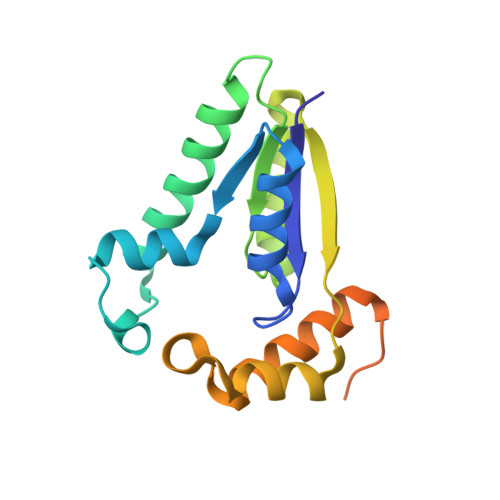
    T4 polynucleotide kinase (Pnk), in addition to being an invaluable research tool, exemplifies a family of bifunctional enzymes with 5'-kinase and 3'-phosphatase activities that play key roles in RNA and DNA repair. T4 Pnk is a homotetramer composed of a C-terminal phosphatase domain and an N-terminal kinase domain. The 2.0 A crystal structure of the isolated kinase domain highlights a tunnel-like active site through the heart of the enzyme, with an entrance on the 5' OH acceptor side that can accommodate a single-stranded polynucleotide. The active site is composed of essential side chains that coordinate the beta phosphate of the NTP donor and the 3' phosphate of the 5' OH acceptor, plus a putative general acid that activates the 5' OH. The structure rationalizes the different specificities of T4 and eukaryotic Pnk and suggests a model for the assembly of the tetramer.


  • Organizational Affiliation

    Molecular Biology Program, Sloan-Kettering Institute, New York, NY 10021, USA.


Macromolecules
Find similar proteins by:  (by identity cutoff)  |  3D Structure
Entity ID: 1
MoleculeChains Sequence LengthOrganismDetailsImage
polynucleotide kinase181Tequatrovirus T4Mutation(s): 0 
Gene Names: PSET
EC: 2.7.1.78
UniProt
Find proteins for P06855 (Enterobacteria phage T4)
Explore P06855 
Go to UniProtKB:  P06855
Entity Groups  
Sequence Clusters30% Identity50% Identity70% Identity90% Identity95% Identity100% Identity
UniProt GroupP06855
Sequence Annotations
Expand
  • Reference Sequence
Experimental Data & Validation

Experimental Data

  • Method: X-RAY DIFFRACTION
  • Resolution: 2.00 Å
  • R-Value Free: 0.231 
  • R-Value Work: 0.210 
  • Space Group: C 2 2 21
Unit Cell:
Length ( Å )Angle ( ˚ )
a = 65.3α = 90
b = 91.8β = 90
c = 67.9γ = 90
Software Package:
Software NamePurpose
SOLVEphasing
CNSrefinement
DENZOdata reduction
SCALEPACKdata scaling

Structure Validation

View Full Validation Report


Entry History 

Deposition Data

Revision History  (Full details and data files)

  • Version 1.0: 2002-07-17
    Type: Initial release
  • Version 1.1: 2008-04-28
    Changes: Version format compliance
  • Version 1.2: 2011-07-13
    Changes: Version format compliance
  • Version 1.3: 2024-02-14
    Changes: Data collection, Database references, Derived calculations