Gram-positive DsbE Proteins Function Differently from Gram-negative DsbE Homologs: A STRUCTURE TO FUNCTION ANALYSIS OF DsbE FROM MYCOBACTERIUM TUBERCULOSIS.
Goulding, C.W., Apostol, M.I., Gleiter, S., Parseghian, A., Bardwell, J., Gennaro, M., Eisenberg, D.(2004) J Biol Chem 279: 3516-3524
- PubMed: 14597624
- DOI: https://doi.org/10.1074/jbc.M311833200
- Primary Citation of Related Structures:
1LU4 - PubMed Abstract:
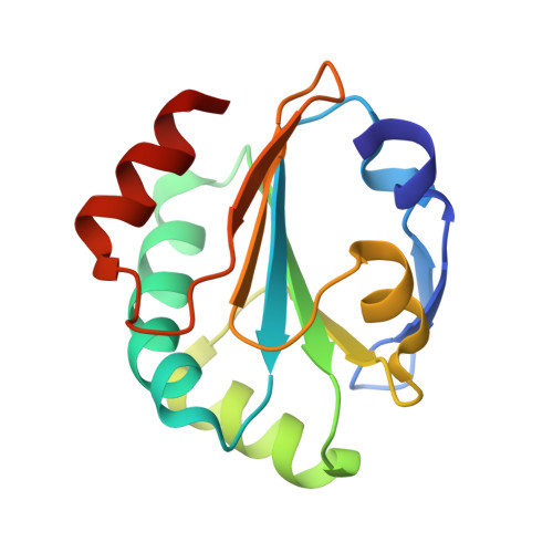
Mycobacterium tuberculosis, a Gram-positive bacterium, encodes a secreted Dsb-like protein annotated as Mtb DsbE (Rv2878c, also known as MPT53). Because Dsb proteins in Escherichia coli and other bacteria seem to catalyze proper folding during protein secretion and because folding of secreted proteins is thought to be coupled to disulfide oxidoreduction, the function of Mtb DsbE may be to ensure that secreted proteins are in their correctly folded states. We have determined the crystal structure of Mtb DsbE to 1.1 A resolution, which reveals a thioredoxin-like domain with a typical CXXC active site. These cysteines are in their reduced state. Biochemical characterization of Mtb DsbE reveals that this disulfide oxidoreductase is an oxidant, unlike Gram-negative bacteria DsbE proteins, which have been shown to be weak reductants. In addition, the pK(a) value of the active site, solvent-exposed cysteine is approximately 2 pH units lower than that of Gram-negative DsbE homologs. Finally, the reduced form of Mtb DsbE is more stable than the oxidized form, and Mtb DsbE is able to oxidatively fold hirudin. Structural and biochemical analysis implies that Mtb DsbE functions differently from Gram-negative DsbE homologs, and we discuss its possible functional role in the bacterium.
Organizational Affiliation:
Howard Hughes Medical Institute and UCLA-Department of Energy Institute of Genomics and Proteomics, Los Angeles, California 90095-1570.