1LRZ

x-ray crystal structure of staphylococcus aureus femA


Experimental Data Snapshot

  • Method: X-RAY DIFFRACTION
  • Resolution: 2.10 Å
  • R-Value Free: 0.212 
  • R-Value Work: 0.180 
  • R-Value Observed: 0.183 

wwPDB Validation   3D Report Full Report


This is version 1.3 of the entry. See complete history


Literature

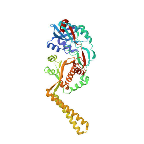
X-ray crystal structure of Staphylococcus aureus FemA.

Benson, T.E.Prince, D.B.Mutchler, V.T.Curry, K.A.Ho, A.M.Sarver, R.W.Hagadorn, J.C.Choi, G.H.Garlick, R.L.

(2002) Structure 10: 1107-1115

  • DOI: https://doi.org/10.1016/s0969-2126(02)00807-9
  • Primary Citation of Related Structures:  
    1LRZ

  • PubMed Abstract: 

    The latter stages of peptidoglycan biosynthesis in Staphylococci involve the synthesis of a pentaglycine bridge on the epsilon amino group of the pentapeptide lysine side chain. Genetic and biochemical evidence suggest that sequential addition of these glycines is catalyzed by three homologous enzymes, FemX (FmhB), FemA, and FemB. The first protein structure from this family, Staphylococcus aureus FemA, has been solved at 2.1 A resolution by X-ray crystallography. The FemA structure reveals a unique organization of several known protein folds involved in peptide and tRNA binding. The surface of the protein also reveals an L-shaped channel suitable for a peptidoglycan substrate. Analysis of the structural features of this enzyme provides clues to the mechanism of action of S. aureus FemA.


  • Organizational Affiliation

    Structural, Analytical, and Medicinal Chemistry, Pharmacia Corporation, Kalamazoo, MI 49007, USA. timothy.e.benson@pharmacia.com


Macromolecules
Find similar proteins by:  (by identity cutoff)  |  3D Structure
Entity ID: 1
MoleculeChains Sequence LengthOrganismDetailsImage
factor essential for expression of methicillin resistance426Staphylococcus aureusMutation(s): 0 
Gene Names: FemA
UniProt
Find proteins for P0A0A5 (Staphylococcus aureus)
Explore P0A0A5 
Go to UniProtKB:  P0A0A5
Entity Groups  
Sequence Clusters30% Identity50% Identity70% Identity90% Identity95% Identity100% Identity
UniProt GroupP0A0A5
Sequence Annotations
Expand
  • Reference Sequence
Experimental Data & Validation

Experimental Data

  • Method: X-RAY DIFFRACTION
  • Resolution: 2.10 Å
  • R-Value Free: 0.212 
  • R-Value Work: 0.180 
  • R-Value Observed: 0.183 
  • Space Group: P 21 21 21
Unit Cell:
Length ( Å )Angle ( ˚ )
a = 53.93α = 90
b = 90.36β = 90
c = 109.35γ = 90
Software Package:
Software NamePurpose
MLPHAREphasing
CNXrefinement
HKL-2000data reduction
SCALEPACKdata scaling

Structure Validation

View Full Validation Report


Entry History 

Deposition Data

Revision History  (Full details and data files)

  • Version 1.0: 2002-09-04
    Type: Initial release
  • Version 1.1: 2008-04-28
    Changes: Version format compliance
  • Version 1.2: 2011-07-13
    Changes: Version format compliance
  • Version 1.3: 2024-02-14
    Changes: Data collection, Database references