1LPA

INTERFACIAL ACTIVATION OF THE LIPASE-PROCOLIPASE COMPLEX BY MIXED MICELLES REVEALED BY X-RAY CRYSTALLOGRAPHY


Experimental Data Snapshot

  • Method: X-RAY DIFFRACTION
  • Resolution: 3.04 Å
  • R-Value Work: 0.186 
  • R-Value Observed: 0.186 

wwPDB Validation   3D Report Full Report


This is version 1.4 of the entry. See complete history


Literature

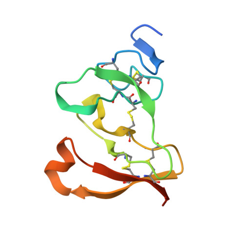
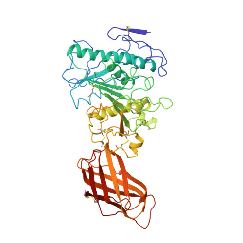
Interfacial activation of the lipase-procolipase complex by mixed micelles revealed by X-ray crystallography.

van Tilbeurgh, H.Egloff, M.P.Martinez, C.Rugani, N.Verger, R.Cambillau, C.

(1993) Nature 362: 814-820

  • DOI: https://doi.org/10.1038/362814a0
  • Primary Citation of Related Structures:  
    1LPA

  • PubMed Abstract: 

    The three-dimensional structure of the lipase-procolipase complex, co-crystallized with mixed micelles of phosphatidylcholine and bile salt, has been determined at 3 A resolution by X-ray crystallography. The lid, a surface helix covering the catalytic triad of lipase, adopts a totally different conformation which allows phospholipid to bind to the enzyme's active site. The open lid is an essential component of the active site and interacts with procolipase. Together they form the lipid-water interface binding site. This reorganization of the lid structure provokes a second drastic conformational change in an active site loop, which in its turn creates the oxyanion hole (induced fit).


  • Organizational Affiliation

    LCCMB-CNRS, Faculté de Médecine Nord, Marseilles, France.


Macromolecules
Find similar proteins by:  (by identity cutoff)  |  3D Structure
Entity ID: 1
MoleculeChains Sequence LengthOrganismDetailsImage
COLIPASE95Sus scrofaMutation(s): 0 
Membrane Entity: Yes 
UniProt
Find proteins for P02703 (Sus scrofa)
Explore P02703 
Go to UniProtKB:  P02703
Entity Groups  
Sequence Clusters30% Identity50% Identity70% Identity90% Identity95% Identity100% Identity
UniProt GroupP02703
Sequence Annotations
Expand
  • Reference Sequence
Find similar proteins by:  (by identity cutoff)  |  3D Structure
Entity ID: 2
MoleculeChains Sequence LengthOrganismDetailsImage
LIPASE449Homo sapiensMutation(s): 0 
EC: 3.1.1.3
Membrane Entity: Yes 
UniProt & NIH Common Fund Data Resources
Find proteins for P16233 (Homo sapiens)
Explore P16233 
Go to UniProtKB:  P16233
PHAROS:  P16233
GTEx:  ENSG00000175535 
Entity Groups  
Sequence Clusters30% Identity50% Identity70% Identity90% Identity95% Identity100% Identity
UniProt GroupP16233
Sequence Annotations
Expand
  • Reference Sequence
Small Molecules
Ligands 3 Unique
IDChains Name / Formula / InChI Key2D Diagram3D Interactions
PLC
Query on PLC

Download Ideal Coordinates CCD File 
E [auth B]DIUNDECYL PHOSPHATIDYL CHOLINE
C32 H65 N O8 P
IJFVSSZAOYLHEE-SSEXGKCCSA-O
BNG
Query on BNG

Download Ideal Coordinates CCD File 
C [auth B]nonyl beta-D-glucopyranoside
C15 H30 O6
QFAPUKLCALRPLH-UXXRCYHCSA-N
CA
Query on CA

Download Ideal Coordinates CCD File 
D [auth B]CALCIUM ION
Ca
BHPQYMZQTOCNFJ-UHFFFAOYSA-N
Experimental Data & Validation

Experimental Data

  • Method: X-RAY DIFFRACTION
  • Resolution: 3.04 Å
  • R-Value Work: 0.186 
  • R-Value Observed: 0.186 
  • Space Group: P 42 21 2
Unit Cell:
Length ( Å )Angle ( ˚ )
a = 133.4α = 90
b = 133.4β = 90
c = 92.6γ = 90
Software Package:
Software NamePurpose
X-PLORmodel building
X-PLORrefinement
X-PLORphasing

Structure Validation

View Full Validation Report


Entry History 

Deposition Data

Revision History  (Full details and data files)

  • Version 1.0: 1994-11-01
    Type: Initial release
  • Version 1.1: 2008-03-03
    Changes: Version format compliance
  • Version 1.2: 2011-07-13
    Changes: Version format compliance
  • Version 1.3: 2017-11-29
    Changes: Derived calculations, Other
  • Version 1.4: 2020-07-29
    Type: Remediation
    Reason: Carbohydrate remediation
    Changes: Data collection, Derived calculations, Structure summary