1LOK

The 1.20 Angstrom Resolution Crystal Structure of the Aminopeptidase from Aeromonas proteolytica Complexed with Tris: A Tale of Buffer Inhibition


Experimental Data Snapshot

  • Method: X-RAY DIFFRACTION
  • Resolution: 1.20 Å
  • R-Value Free: 0.198 
  • R-Value Work: 0.158 
  • R-Value Observed: 0.145 

wwPDB Validation   3D Report Full Report


This is version 1.4 of the entry. See complete history


Literature

The 1.20 Angstrom Resolution Crystal Structure of the Aminopeptidase from Aeromonas proteolytica Complexed with Tris A tale of Buffer Inhibition

Desmarais, W.T.Bienvenue, D.L.Bzymek, K.P.Holz, R.C.Petsko, G.A.Ringe, D.

(2002) Structure 10: 1063-1072

  • DOI: https://doi.org/10.1016/s0969-2126(02)00810-9
  • Primary Citation of Related Structures:  
    1LOK

  • PubMed Abstract: 

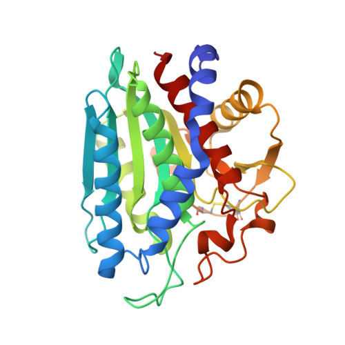
    The aminopeptidase from Aeromonas proteolytica (AAP) is a bridged bimetallic enzyme that removes the N-terminal amino acid from a peptide chain. To fully understand the metal roles in the reaction pathway of AAP we have solved the 1.20 A resolution crystal structure of native AAP (PDB ID = 1LOK). The high-quality electron density maps showed a single Tris molecule chelated to the active site Zn(2+), alternate side chain conformations for some side chains, a sodium ion that mediates a crystal contact, a surface thiocyanate ion, and several potential hydrogen atoms. In addition, the high precision of the atomic positions has led to insight into the protonation states of some of the active site amino acid side chains.


  • Organizational Affiliation

    Program in Biophysics and Structural Biology, Brandeis University, Waltham, Massachusetts 02454, USA.


Macromolecules
Find similar proteins by:  (by identity cutoff)  |  3D Structure
Entity ID: 1
MoleculeChains Sequence LengthOrganismDetailsImage
Bacterial leucyl aminopeptidase291Vibrio proteolyticusMutation(s): 0 
EC: 3.4.11.10
UniProt
Find proteins for Q01693 (Vibrio proteolyticus)
Explore Q01693 
Go to UniProtKB:  Q01693
Entity Groups  
Sequence Clusters30% Identity50% Identity70% Identity90% Identity95% Identity100% Identity
UniProt GroupQ01693
Sequence Annotations
Expand
  • Reference Sequence
Small Molecules
Ligands 4 Unique
IDChains Name / Formula / InChI Key2D Diagram3D Interactions
TRS
Query on TRS

Download Ideal Coordinates CCD File 
F [auth A]2-AMINO-2-HYDROXYMETHYL-PROPANE-1,3-DIOL
C4 H12 N O3
LENZDBCJOHFCAS-UHFFFAOYSA-O
ZN
Query on ZN

Download Ideal Coordinates CCD File 
B [auth A],
C [auth A]
ZINC ION
Zn
PTFCDOFLOPIGGS-UHFFFAOYSA-N
SCN
Query on SCN

Download Ideal Coordinates CCD File 
E [auth A]THIOCYANATE ION
C N S
ZMZDMBWJUHKJPS-UHFFFAOYSA-M
NA
Query on NA

Download Ideal Coordinates CCD File 
D [auth A]SODIUM ION
Na
FKNQFGJONOIPTF-UHFFFAOYSA-N
Experimental Data & Validation

Experimental Data

  • Method: X-RAY DIFFRACTION
  • Resolution: 1.20 Å
  • R-Value Free: 0.198 
  • R-Value Work: 0.158 
  • R-Value Observed: 0.145 
  • Space Group: P 61 2 2
Unit Cell:
Length ( Å )Angle ( ˚ )
a = 108.338α = 90
b = 108.338β = 90
c = 93.519γ = 120
Software Package:
Software NamePurpose
SHELXL-97refinement
SCALEPACKdata scaling
CNSrefinement
DENZOdata reduction
CNSphasing

Structure Validation

View Full Validation Report


Entry History 

Deposition Data

Revision History  (Full details and data files)

  • Version 1.0: 2002-11-27
    Type: Initial release
  • Version 1.1: 2008-04-28
    Changes: Version format compliance
  • Version 1.2: 2011-07-13
    Changes: Version format compliance
  • Version 1.3: 2019-07-24
    Changes: Data collection, Refinement description
  • Version 1.4: 2023-08-16
    Changes: Data collection, Database references, Derived calculations, Refinement description