1LOG

X-RAY STRUCTURE OF A (ALPHA-MAN(1-3)BETA-MAN(1-4)GLCNAC)-LECTIN COMPLEX AT 2.1 ANGSTROMS RESOLUTION


Experimental Data Snapshot

  • Method: X-RAY DIFFRACTION
  • Resolution: 2.10 Å
  • R-Value Work: 0.175 
  • R-Value Observed: 0.175 

wwPDB Validation   3D Report Full Report


This is version 2.1 of the entry. See complete history


Literature

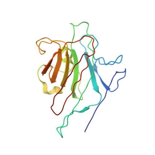
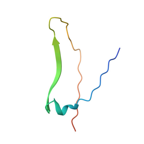
X-ray structure of a (alpha-Man(1-3)beta-Man(1-4)GlcNAc)-lectin complex at 2.1-A resolution. The role of water in sugar-lectin interaction.

Bourne, Y.Rouge, P.Cambillau, C.

(1990) J Biol Chem 265: 18161-18165

  • Primary Citation of Related Structures:  
    1LOG

  • PubMed Abstract: 

    We describe herein the high resolution refined x-ray structure of a trisaccharide, which is a part of the N-acetyllactosamine type glycan found in the majority of the N-glycosyl-proteins, complexed to the isolectin I. According to the potentials used by Imberty et al. (Imburty, A., Gerber, S., Tran, V., and Pérez, S. (1990) Glycoconjugate J. 7, 27-54) the trisaccharide is in a low-energy state. Only one mannose moiety establishes direct hydrogen bonds with the lectin, as it is the case for monosaccharide-lectin complexes. The comparison of our trisaccharide with the one determined in solution by Warin et al. (Warin, V., Baert, F., Fouret, R., Strecker, G., Fournet, B., and Montreuil, J. (1979) Carbohydr. Res. 76, 11-22) shows that both adopt roughly the same conformation. The differences in these two sugar structures allow us to assign the role of water molecules present in the vicinity of our trisaccharide for the stabilization of this sugar-lectin complex.


  • Organizational Affiliation

    Laboratoire de Cristallographie et de Cristallisation des Macromolécules Biologiques, Faculté de Médecine, Marseille, France.


Macromolecules
Find similar proteins by:  (by identity cutoff)  |  3D Structure
Entity ID: 1
MoleculeChains Sequence LengthOrganismDetailsImage
LEGUME ISOLECTIN I (ALPHA CHAIN)
A, C
181Lathyrus ochrusMutation(s): 0 
UniProt
Find proteins for P04122 (Lathyrus ochrus)
Explore P04122 
Go to UniProtKB:  P04122
Entity Groups  
Sequence Clusters30% Identity50% Identity70% Identity90% Identity95% Identity100% Identity
UniProt GroupP04122
Sequence Annotations
Expand
  • Reference Sequence
Find similar proteins by:  (by identity cutoff)  |  3D Structure
Entity ID: 2
MoleculeChains Sequence LengthOrganismDetailsImage
LEGUME ISOLECTIN I (BETA CHAIN)
B, D
52Lathyrus ochrusMutation(s): 0 
UniProt
Find proteins for P12306 (Lathyrus ochrus)
Explore P12306 
Go to UniProtKB:  P12306
Entity Groups  
Sequence Clusters30% Identity50% Identity70% Identity90% Identity95% Identity100% Identity
UniProt GroupP12306
Sequence Annotations
Expand
  • Reference Sequence
Oligosaccharides

Help

Entity ID: 3
MoleculeChains Length2D Diagram Glycosylation3D Interactions
alpha-D-mannopyranose-(1-3)-beta-D-mannopyranose-(1-4)-2-acetamido-2-deoxy-beta-D-glucopyranose
E, F
3N/A
Glycosylation Resources
GlyTouCan:  G29301NP
GlyCosmos:  G29301NP
GlyGen:  G29301NP
Experimental Data & Validation

Experimental Data

  • Method: X-RAY DIFFRACTION
  • Resolution: 2.10 Å
  • R-Value Work: 0.175 
  • R-Value Observed: 0.175 
  • Space Group: P 21 21 2
Unit Cell:
Length ( Å )Angle ( ˚ )
a = 137.1α = 90
b = 63.3β = 90
c = 54.6γ = 90
Software Package:
Software NamePurpose
X-PLORmodel building
X-PLORrefinement
X-PLORphasing

Structure Validation

View Full Validation Report


Entry History 

Deposition Data

Revision History  (Full details and data files)

  • Version 1.0: 1994-04-30
    Type: Initial release
  • Version 1.1: 2008-03-24
    Changes: Version format compliance
  • Version 1.2: 2011-07-13
    Changes: Derived calculations, Version format compliance
  • Version 2.0: 2020-07-29
    Type: Remediation
    Reason: Carbohydrate remediation
    Changes: Atomic model, Data collection, Database references, Derived calculations, Other, Structure summary
  • Version 2.1: 2024-02-14
    Changes: Data collection, Database references, Structure summary