1LOF

X-RAY STRUCTURE OF A BIANTENNARY OCTASACCHARIDE-LECTIN COMPLEX AT 2.3 ANGSTROMS RESOLUTION


Experimental Data Snapshot

  • Method: X-RAY DIFFRACTION
  • Resolution: 2.30 Å
  • R-Value Work: 0.190 
  • R-Value Observed: 0.190 

wwPDB Validation   3D Report Full Report


This is version 2.1 of the entry. See complete history


Literature

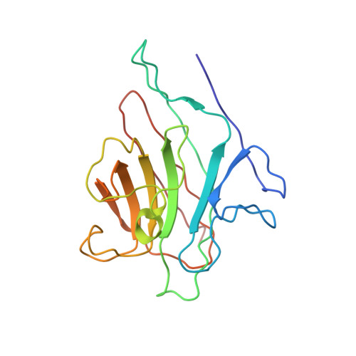
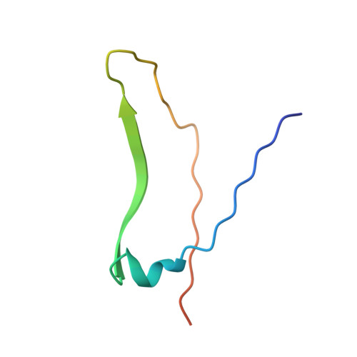
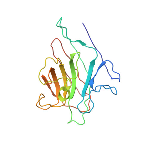
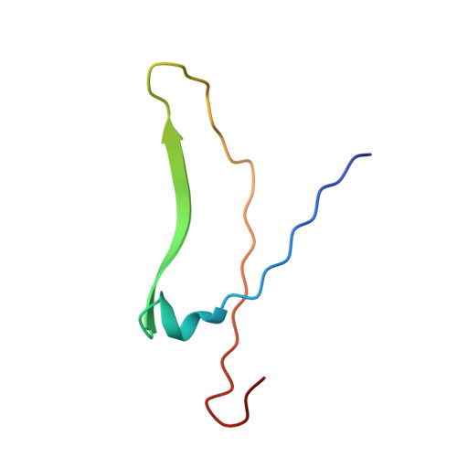
X-ray structure of a biantennary octasaccharide-lectin complex refined at 2.3-A resolution.

Bourne, Y.Rouge, P.Cambillau, C.

(1992) J Biol Chem 267: 197-203

  • Primary Citation of Related Structures:  
    1LOF

  • PubMed Abstract: 

    The structure and flexibility of saccharides have a profound and specific influence in several biological processes such as protein protection and the maintenance of conformational integrity, and in recognition events involving viruses, enzymes, and lectins. To establish the structural bases of these phenomena, we describe herein the extensively refined 2.3-A resolution x-ray structure of a biantennary octasaccharide of the N-acetyllactosamine type, complexed to isolectin I from Lathyrus ochrus. The two octasaccharides are located in clefts at each end of the long axis of the lectin. The conformations of both the lectin and the saccharide are slightly modified upon binding. The complex is stabilized by numerous hydrogen bonds, many of them involving water molecules. It is also stabilized by van der Waals interactions, including some with aromatic residues. A more general model of a possible lectin-glycoprotein interaction is also proposed.


  • Organizational Affiliation

    Laboratoire de Cristallographie et de Cristallisation des Macromolécules Biologiques, Faculté de Médecine Secteur-Nord, Marseille, France.


Macromolecules
Find similar proteins by:  (by identity cutoff)  |  3D Structure
Entity ID: 1
MoleculeChains Sequence LengthOrganismDetailsImage
LEGUME ISOLECTIN I (ALPHA CHAIN)181Lathyrus ochrusMutation(s): 0 
UniProt
Find proteins for P04122 (Lathyrus ochrus)
Explore P04122 
Go to UniProtKB:  P04122
Entity Groups  
Sequence Clusters30% Identity50% Identity70% Identity90% Identity95% Identity100% Identity
UniProt GroupP04122
Sequence Annotations
Expand
  • Reference Sequence
Find similar proteins by:  (by identity cutoff)  |  3D Structure
Entity ID: 2
MoleculeChains Sequence LengthOrganismDetailsImage
LEGUME ISOLECTIN I (BETA CHAIN)52Lathyrus ochrusMutation(s): 0 
UniProt
Find proteins for P12306 (Lathyrus ochrus)
Explore P12306 
Go to UniProtKB:  P12306
Entity Groups  
Sequence Clusters30% Identity50% Identity70% Identity90% Identity95% Identity100% Identity
UniProt GroupP12306
Sequence Annotations
Expand
  • Reference Sequence
Find similar proteins by:  (by identity cutoff)  |  3D Structure
Entity ID: 3
MoleculeChains Sequence LengthOrganismDetailsImage
LEGUME ISOLECTIN I (ALPHA CHAIN)181Lathyrus ochrusMutation(s): 0 
UniProt
Find proteins for P04122 (Lathyrus ochrus)
Explore P04122 
Go to UniProtKB:  P04122
Entity Groups  
Sequence Clusters30% Identity50% Identity70% Identity90% Identity95% Identity100% Identity
UniProt GroupP04122
Sequence Annotations
Expand
  • Reference Sequence
Find similar proteins by:  (by identity cutoff)  |  3D Structure
Entity ID: 4
MoleculeChains Sequence LengthOrganismDetailsImage
LEGUME ISOLECTIN I (BETA CHAIN)52Lathyrus ochrusMutation(s): 0 
UniProt
Find proteins for P12306 (Lathyrus ochrus)
Explore P12306 
Go to UniProtKB:  P12306
Entity Groups  
Sequence Clusters30% Identity50% Identity70% Identity90% Identity95% Identity100% Identity
UniProt GroupP12306
Sequence Annotations
Expand
  • Reference Sequence
Oligosaccharides

Help

Entity ID: 5
MoleculeChains Length2D Diagram Glycosylation3D Interactions
beta-D-galactopyranose-(1-4)-2-acetamido-2-deoxy-beta-D-glucopyranose-(1-2)-alpha-D-mannopyranose-(1-3)-[beta-D-galactopyranose-(1-4)-2-acetamido-2-deoxy-beta-D-glucopyranose-(1-2)-alpha-D-mannopyranose-(1-6)]beta-D-mannopyranose-(1-4)-2-acetamido-2-deoxy-beta-D-glucopyranose
E
8N/A
Glycosylation Resources
GlyTouCan:  G41327HW
GlyCosmos:  G41327HW
GlyGen:  G41327HW
Entity ID: 6
MoleculeChains Length2D Diagram Glycosylation3D Interactions
2-acetamido-2-deoxy-beta-D-glucopyranose-(1-2)-alpha-D-mannopyranose-(1-3)-alpha-D-mannopyranose
F
3N/A
Glycosylation Resources
GlyTouCan:  G57278AJ
GlyCosmos:  G57278AJ
GlyGen:  G57278AJ
Experimental Data & Validation

Experimental Data

  • Method: X-RAY DIFFRACTION
  • Resolution: 2.30 Å
  • R-Value Work: 0.190 
  • R-Value Observed: 0.190 
  • Space Group: C 1 2 1
Unit Cell:
Length ( Å )Angle ( ˚ )
a = 78.3α = 90
b = 75.4β = 92
c = 103.9γ = 90
Software Package:
Software NamePurpose
X-PLORmodel building
X-PLORrefinement
X-PLORphasing

Structure Validation

View Full Validation Report


Entry History 

Deposition Data

Revision History  (Full details and data files)

  • Version 1.0: 1994-04-30
    Type: Initial release
  • Version 1.1: 2008-03-24
    Changes: Version format compliance
  • Version 1.2: 2011-07-13
    Changes: Derived calculations, Version format compliance
  • Version 2.0: 2020-07-29
    Type: Remediation
    Reason: Carbohydrate remediation
    Changes: Advisory, Atomic model, Data collection, Database references, Derived calculations, Other, Structure summary
  • Version 2.1: 2024-02-14
    Changes: Data collection, Database references, Structure summary