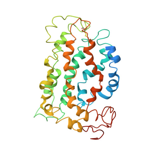
1LN6

STRUCTURE OF BOVINE RHODOPSIN (Metarhodopsin II)


Experimental Data Snapshot

  • Method: SOLUTION NMR
  • Conformers Calculated: 10 
  • Conformers Submitted: 
  • Selection Criteria: structures with the lowest energy 

wwPDB Validation   3D Report Full Report


This is version 1.3 of the entry. See complete history


Literature

Structural studies of metarhodopsin II, the activated form of the G-protein coupled receptor, rhodopsin.

Choi, G.Landin, J.Galan, J.F.Birge, R.R.Albert, A.D.Yeagle, P.L.

(2002) Biochemistry 41: 7318-7324

  • DOI: https://doi.org/10.1021/bi025507w
  • Primary Citation of Related Structures:  
    1LN6

  • PubMed Abstract: 

    The structural changes that accompany activation of a G-protein coupled receptor (GPCR) are not well understood. To better understand the activation of rhodopsin, the GPCR responsible for visual transduction, we report studies on the three-dimensional structure for the activated state of this receptor, metarhodopsin II. Differences between the three-dimensional structure of ground state rhodopsin and metarhodopsin II, particularly in the cytoplasmic face of the receptor, suggest how the receptor is activated to couple with transducin. In particular, activation opens a groove on the surface of the receptor that could bind the N-terminal helix of the G protein, transducin alpha.


  • Organizational Affiliation

    Department of Molecular and Cell Biology, University of Connecticut, Storrs, CT 06269, USA.


Macromolecules
Find similar proteins by:  (by identity cutoff)  |  3D Structure
Entity ID: 1
MoleculeChains Sequence LengthOrganismDetailsImage
RHODOPSIN348N/AMutation(s): 0 
Membrane Entity: Yes 
UniProt
Find proteins for P02699 (Bos taurus)
Explore P02699 
Go to UniProtKB:  P02699
Entity Groups  
Sequence Clusters30% Identity50% Identity70% Identity90% Identity95% Identity100% Identity
UniProt GroupP02699
Sequence Annotations
Expand
  • Reference Sequence
Small Molecules
Ligands 1 Unique
IDChains Name / Formula / InChI Key2D Diagram3D Interactions
RET
Query on RET

Download Ideal Coordinates CCD File 
B [auth A]RETINAL
C20 H28 O
NCYCYZXNIZJOKI-OVSJKPMPSA-N
Experimental Data & Validation

Experimental Data

  • Method: SOLUTION NMR
  • Conformers Calculated: 10 
  • Conformers Submitted: 
  • Selection Criteria: structures with the lowest energy 

Structure Validation

View Full Validation Report


Entry History 

Deposition Data

Revision History  (Full details and data files)

  • Version 1.0: 2002-07-10
    Type: Initial release
  • Version 1.1: 2008-04-28
    Changes: Version format compliance
  • Version 1.2: 2011-07-13
    Changes: Version format compliance
  • Version 1.3: 2022-02-23
    Changes: Database references, Derived calculations