1LKD

CRYSTAL STRUCTURE OF 2,3-DIHYDROXYBIPHENYL 1,2-DIOXYGENASE (DHBD) COMPLEXED WITH 2',6'-DICL DIHYDROXYBIPHENYL (DHB)


Experimental Data Snapshot

  • Method: X-RAY DIFFRACTION
  • Resolution: 1.70 Å
  • R-Value Free: 0.220 
  • R-Value Work: 0.203 

wwPDB Validation   3D Report Full Report


Ligand Structure Quality Assessment 


This is version 1.3 of the entry. See complete history


Literature

Identification and analysis of a bottleneck in PCB biodegradation

Dai, S.Vaillancourt, F.H.Maaroufi, H.Drouin, N.M.Neau, D.B.Snieckus, V.Bolin, J.T.Eltis, L.D.

(2002) Nat Struct Biol 9: 934-939

  • DOI: https://doi.org/10.1038/nsb866
  • Primary Citation of Related Structures:  
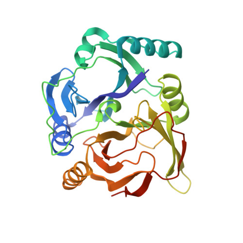
    1LGT, 1LKD

  • PubMed Abstract: 

    The microbial degradation of polychlorinated biphenyls (PCBs) provides the potential to destroy these widespread, toxic and persistent environmental pollutants. For example, the four-step upper bph pathway transforms some of the more than 100 different PCBs found in commercial mixtures and is being engineered for more effective PCB degradation. In the critical third step of this pathway, 2,3-dihydroxybiphenyl (DHB) 1,2-dioxygenase (DHBD; EC 1.13.11.39) catalyzes aromatic ring cleavage. Here we demonstrate that ortho-chlorinated PCB metabolites strongly inhibit DHBD, promote its suicide inactivation and interfere with the degradation of other compounds. For example, k(cat)(app) for 2',6'-diCl DHB was reduced by a factor of approximately 7,000 relative to DHB, and it bound with sufficient affinity to competitively inhibit DHB cleavage at nanomolar concentrations. Crystal structures of two complexes of DHBD with ortho-chlorinated metabolites at 1.7 A resolution reveal an explanation for these phenomena, which have important implications for bioremediation strategies.


  • Organizational Affiliation

    Markey Center for Structural Biology, Department of Biological Sciences, Purdue University, West Lafayette, Indiana 47907-1392, USA.


Macromolecules
Find similar proteins by:  (by identity cutoff)  |  3D Structure
Entity ID: 1
MoleculeChains Sequence LengthOrganismDetailsImage
BIPHENYL-2,3-DIOL 1,2-DIOXYGENASE297Paraburkholderia xenovorans LB400Mutation(s): 0 
EC: 1.13.11.39
UniProt
Find proteins for P47228 (Paraburkholderia xenovorans (strain LB400))
Explore P47228 
Go to UniProtKB:  P47228
Entity Groups  
Sequence Clusters30% Identity50% Identity70% Identity90% Identity95% Identity100% Identity
UniProt GroupP47228
Sequence Annotations
Expand
  • Reference Sequence
Experimental Data & Validation

Experimental Data

  • Method: X-RAY DIFFRACTION
  • Resolution: 1.70 Å
  • R-Value Free: 0.220 
  • R-Value Work: 0.203 
  • Space Group: I 4 2 2
Unit Cell:
Length ( Å )Angle ( ˚ )
a = 122.64α = 90
b = 122.64β = 90
c = 107.232γ = 90
Software Package:
Software NamePurpose
HKL-2000data collection
SCALEPACKdata scaling
AMoREphasing
CNSrefinement
HKL-2000data reduction

Structure Validation

View Full Validation Report


Ligand Structure Quality Assessment 


Entry History 

Deposition Data

Revision History  (Full details and data files)

  • Version 1.0: 2002-11-27
    Type: Initial release
  • Version 1.1: 2008-04-28
    Changes: Version format compliance
  • Version 1.2: 2011-07-13
    Changes: Derived calculations, Source and taxonomy, Version format compliance
  • Version 1.3: 2024-02-14
    Changes: Data collection, Database references, Derived calculations