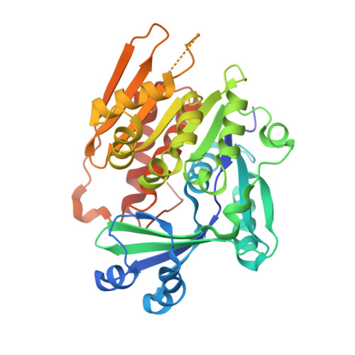
Crystal structures of Toxoplasma gondii adenosine kinase reveal a novel catalytic mechanism and prodrug binding.
Schumacher, M.A., Scott, D.M., Mathews, I.I., Ealick, S.E., Roos, D.S., Ullman, B., Brennan, R.G.(2000) J Mol Biol 298: 875-893
- PubMed: 10801355
- DOI: https://doi.org/10.1006/jmbi.2000.3753
- Primary Citation of Related Structures:
1LII, 1LIJ, 1LIK, 1LIO - PubMed Abstract:
Adenosine kinase (AK) is a key purine metabolic enzyme from the opportunistic parasitic protozoan Toxoplasma gondii and belongs to the family of carbohydrate kinases that includes ribokinase. To understand the catalytic mechanism of AK, we determined the structures of the T. gondii apo AK, AK:adenosine complex and the AK:adenosine:AMP-PCP complex to 2.55 A, 2.50 A and 1.71 A resolution, respectively. These structures reveal a novel catalytic mechanism that involves an adenosine-induced domain rotation of 30 degrees and a newly described anion hole (DTXGAGD), requiring a helix-to-coil conformational change that is induced by ATP binding. Nucleotide binding also evokes a coil-to-helix transition that completes the formation of the ATP binding pocket. A conserved dipeptide, Gly68-Gly69, which is located at the bottom of the adenosine-binding site, functions as the switch for domain rotation. The synergistic structural changes that occur upon substrate binding sequester the adenosine and the ATP gamma phosphate from solvent and optimally position the substrates for catalysis. Finally, the 1.84 A resolution structure of an AK:7-iodotubercidin:AMP-PCP complex reveals the basis for the higher affinity binding of this prodrug over adenosine and thus provides a scaffold for the design of new inhibitors and subversive substrates that target the T. gondii AK.
Organizational Affiliation:
Department of Biochemistry and Molecular Biology, Vollum Institute, Oregon Health Sciences University, Portland 97201-3098, USA.