1LGY

LIPASE II FROM RHIZOPUS NIVEUS


Experimental Data Snapshot

  • Method: X-RAY DIFFRACTION
  • Resolution: 2.20 Å
  • R-Value Work: 0.186 

wwPDB Validation   3D Report Full Report


This is version 1.2 of the entry. See complete history


Literature

The crystal structure of lipase II from Rhizopus niveus at 2.2 A resolution.

Kohno, M.Funatsu, J.Mikami, B.Kugimiya, W.Matsuo, T.Morita, Y.

(1996) J Biochem 120: 505-510

  • DOI: https://doi.org/10.1093/oxfordjournals.jbchem.a021442
  • Primary Citation of Related Structures:  
    1LGY

  • PubMed Abstract: 

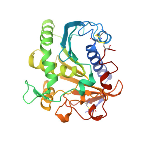
    The crystal and molecular structure of Lipase II from Rhizopus niveus was analyzed using X-ray single crystal diffraction data at a resolution of 2.2 A. The structure was refined to an R-factor of 0.19 for all available data. This lipase was purified and crystallized as Lipase I, which contains two polypeptide chains combined through non-covalent interaction. However, during crystal growth, Lipase I was converted to Lipase II, which consists of a single polypeptide chain of 269 amino acid residues, by limited proteolysis. The structure of Lipase II shows a typical alpha/beta hydrolase fold containing the so-called nucleophilic elbow. The catalytic center of this enzyme is analogous to those of other neutral lipases and serine proteases. This catalytic center is sheltered by an alpha-helix lid, which appears in neutral lipases, opening the active site at the oil-water interface.


  • Organizational Affiliation

    Central Research Institute Tsukuba R&D Center, Fuji Oil Co., Ltd., Ibaraki. kouno@fujioil.co.jp


Macromolecules
Find similar proteins by:  (by identity cutoff)  |  3D Structure
Entity ID: 1
MoleculeChains Sequence LengthOrganismDetailsImage
TRIACYLGLYCEROL LIPASE
A, B, C
269Rhizopus niveusMutation(s): 0 
EC: 3.1.1.3
Membrane Entity: Yes 
UniProt
Find proteins for P61871 (Rhizopus niveus)
Explore P61871 
Go to UniProtKB:  P61871
Entity Groups  
Sequence Clusters30% Identity50% Identity70% Identity90% Identity95% Identity100% Identity
UniProt GroupP61871
Sequence Annotations
Expand
  • Reference Sequence
Experimental Data & Validation

Experimental Data

  • Method: X-RAY DIFFRACTION
  • Resolution: 2.20 Å
  • R-Value Work: 0.186 
  • Space Group: P 43
Unit Cell:
Length ( Å )Angle ( ˚ )
a = 83.7α = 90
b = 83.7β = 90
c = 137.9γ = 90
Software Package:
Software NamePurpose
RIGAKUdata collection
X-PLORmodel building
TNTrefinement
X-PLORrefinement
RIGAKUdata reduction
X-PLORphasing

Structure Validation

View Full Validation Report


Entry History 

Deposition Data

Revision History  (Full details and data files)

  • Version 1.0: 1996-12-23
    Type: Initial release
  • Version 1.1: 2008-03-24
    Changes: Version format compliance
  • Version 1.2: 2011-07-13
    Changes: Version format compliance