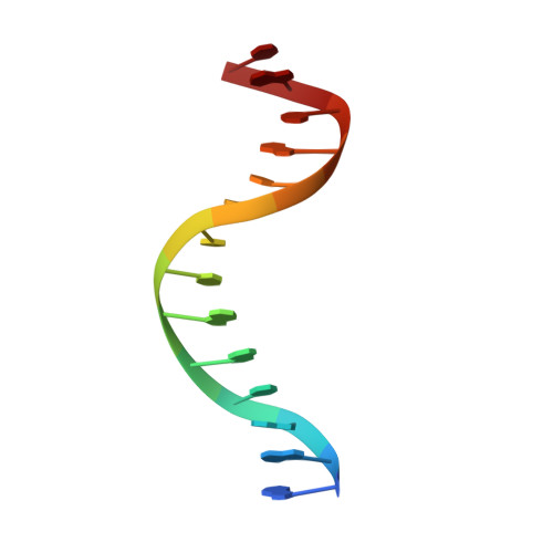
1LFU

NMR Solution Structure of the Extended PBX Homeodomain Bound to DNA


Experimental Data Snapshot

  • Method: SOLUTION NMR
  • Conformers Calculated: 100 
  • Conformers Submitted: 20 
  • Selection Criteria: lowest energy with acceptable geometry 

wwPDB Validation   3D Report Full Report


This is version 1.4 of the entry. See complete history


Literature

Lock and Key Binding of the HOX YPWM Peptide to the PBX Homeodomain

Sprules, T.Green, N.Featherstone, M.Gehring, K.

(2003) J Biol Chem 278: 1053-1058

  • DOI: https://doi.org/10.1074/jbc.M207504200
  • Primary Citation of Related Structures:  
    1LFU

  • PubMed Abstract: 

    HOX homeodomain proteins bind short core DNA sequences to control very specific developmental processes. DNA binding affinity and sequence selectivity are increased by the formation of cooperative complexes with the PBX homeodomain protein. A conserved YPWM motif in the HOX protein is necessary for cooperative binding with PBX. We have determined the structure of a PBX homeodomain bound to a 14-mer DNA duplex. A relaxation-optimized procedure was developed to measure DNA residual dipolar couplings at natural abundance in the 20-kDa binary complex. When the PBX homeodomain binds to DNA, a fourth alpha-helix is formed in the homeodomain. This helix rigidifies the DNA recognition helix of PBX and forms a hydrophobic binding site for the HOX YPWM peptide. The HOX peptide itself shows some structure in solution and suggests that the interaction between PBX and HOX is an example of "lock and key" binding. The NMR structure explains the requirement of DNA for the PBX-HOX interaction and the increased affinity of DNA binding.


  • Organizational Affiliation

    Department of Biochemistry, McGill University, Montreal, Quebec H3G 1Y6, Canada.


Macromolecules

Find similar proteins by:  (by identity cutoff)  |  3D Structure
Entity ID: 3
MoleculeChains Sequence LengthOrganismDetailsImage
homeobox protein PBX1C [auth P]82Mus musculusMutation(s): 1 
Gene Names: PBX1
UniProt
Find proteins for P41778 (Mus musculus)
Explore P41778 
Go to UniProtKB:  P41778
Entity Groups  
Sequence Clusters30% Identity50% Identity70% Identity90% Identity95% Identity100% Identity
UniProt GroupP41778
Sequence Annotations
Expand
  • Reference Sequence

Find similar nucleic acids by:  Sequence   |   3D Structure  

Entity ID: 1
MoleculeChains LengthOrganismImage
5'-D(*GP*CP*GP*CP*AP*TP*GP*AP*TP*TP*GP*CP*CP*C)-3'14N/A
Sequence Annotations
Expand
  • Reference Sequence

Find similar nucleic acids by:  Sequence   |   3D Structure  

Entity ID: 2
MoleculeChains LengthOrganismImage
5'-D(*GP*GP*GP*CP*AP*AP*TP*CP*AP*TP*GP*CP*GP*C)-3'14N/A
Sequence Annotations
Expand
  • Reference Sequence
Experimental Data & Validation

Experimental Data

  • Method: SOLUTION NMR
  • Conformers Calculated: 100 
  • Conformers Submitted: 20 
  • Selection Criteria: lowest energy with acceptable geometry 

Structure Validation

View Full Validation Report


Entry History 

Deposition Data

Revision History  (Full details and data files)

  • Version 1.0: 2003-01-14
    Type: Initial release
  • Version 1.1: 2008-04-28
    Changes: Version format compliance
  • Version 1.2: 2011-07-13
    Changes: Version format compliance
  • Version 1.3: 2017-02-01
    Changes: Structure summary
  • Version 1.4: 2021-10-27
    Changes: Data collection, Database references