1LFO

LIVER FATTY ACID BINDING PROTEIN-OLEATE COMPLEX


Experimental Data Snapshot

  • Method: X-RAY DIFFRACTION
  • Resolution: 2.30 Å
  • R-Value Free: 0.262 
  • R-Value Work: 0.202 
  • R-Value Observed: 0.202 

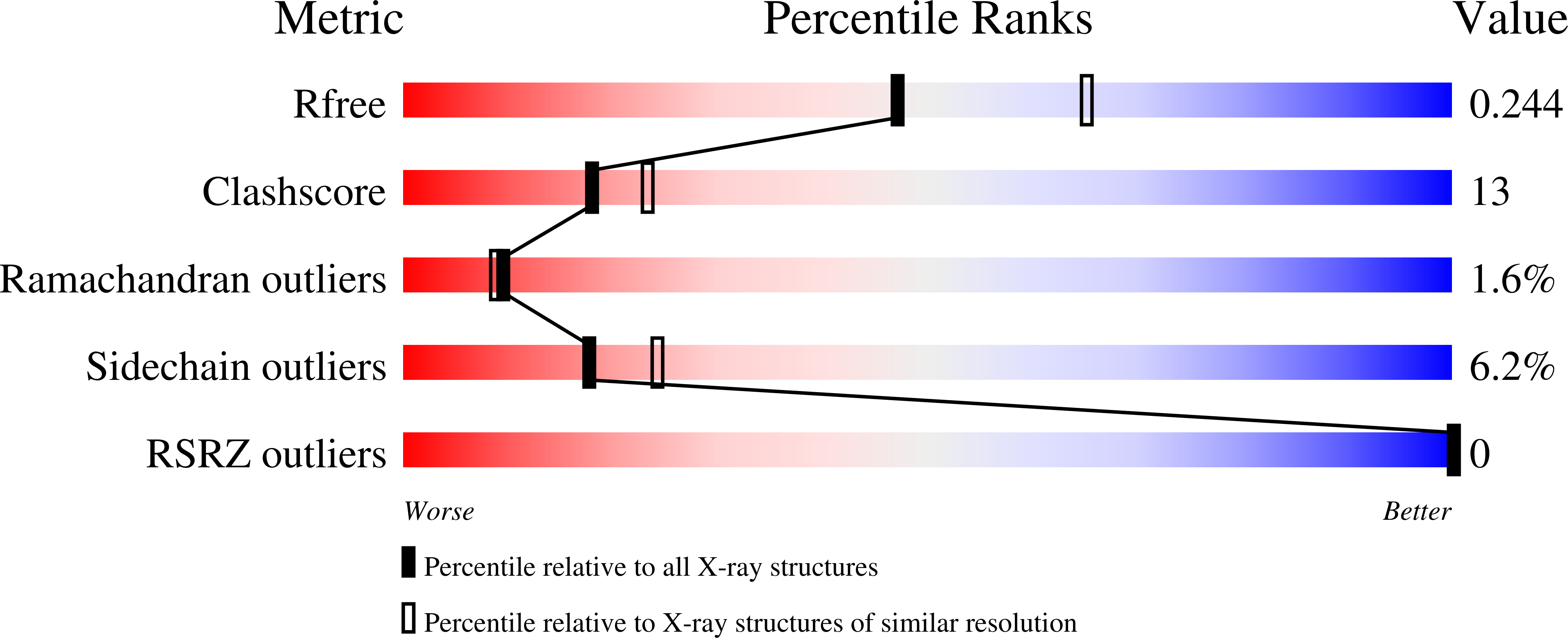
wwPDB Validation   3D Report Full Report


Ligand Structure Quality Assessment 


This is version 1.3 of the entry. See complete history


Literature

The crystal structure of the liver fatty acid-binding protein. A complex with two bound oleates.

Thompson, J.Winter, N.Terwey, D.Bratt, J.Banaszak, L.

(1997) J Biol Chem 272: 7140-7150

  • DOI: https://doi.org/10.1074/jbc.272.11.7140
  • Primary Citation of Related Structures:  
    1LFO

  • PubMed Abstract: 

    The crystal structure of the recombinant form of rat liver fatty acid-binding protein was completed to 2.3 A and refined to an R factor of 19.0%. The structural solution was obtained by molecular replacement using superimposed polyalanine coordinates of six intracellular lipid-binding proteins as a search probe. The entire amino acid sequence of rat liver fatty acid-binding protein along with an amino-terminal formyl-methionine was modeled in the crystal structure. In addition, the crystal was obtained in the presence of oleic acid, and the initial electron density clearly showed two fatty acid molecules bound within a central cavity. The carboxylate of one fatty acid molecule interacts with arginine 122 and is shielded from free solvent. It has an overall bent conformation. The more solvent-exposed carboxylate of the other oleate is located near the helix-turn-helix that caps one end of the beta-barrel, while the acyl chain lies in the interior. The cavity contains both polar and nonpolar residues but also shows extensive hydrophobic character around the nonpolar atoms of the ligands. The primary and secondary oleate binding sites appear to be totally interdependent, mainly because favorable hydrophobic interactions form between both aliphatic chains.


  • Organizational Affiliation

    Department of Biochemistry, University of Minnesota Medical School, Minneapolis, Minnesota 55455, USA.


Macromolecules
Find similar proteins by:  (by identity cutoff)  |  3D Structure
Entity ID: 1
MoleculeChains Sequence LengthOrganismDetailsImage
LIVER FATTY ACID BINDING PROTEIN128Rattus norvegicusMutation(s): 1 
UniProt
Find proteins for P02692 (Rattus norvegicus)
Explore P02692 
Go to UniProtKB:  P02692
Entity Groups  
Sequence Clusters30% Identity50% Identity70% Identity90% Identity95% Identity100% Identity
UniProt GroupP02692
Sequence Annotations
Expand
  • Reference Sequence
Small Molecules
Modified Residues  1 Unique
IDChains TypeFormula2D DiagramParent
SMC
Query on SMC
A
L-PEPTIDE LINKINGC4 H9 N O2 SCYS
Binding Affinity Annotations 
IDSourceBinding Affinity
OLA BindingDB:  1LFO Ki: min: 180, max: 2900 (nM) from 2 assay(s)
PDBBind:  1LFO Kd: 200 (nM) from 1 assay(s)
Binding MOAD:  1LFO Kd: 2000 (nM) from 1 assay(s)
Experimental Data & Validation

Experimental Data

  • Method: X-RAY DIFFRACTION
  • Resolution: 2.30 Å
  • R-Value Free: 0.262 
  • R-Value Work: 0.202 
  • R-Value Observed: 0.202 
  • Space Group: P 32 2 1
Unit Cell:
Length ( Å )Angle ( ˚ )
a = 83.89α = 90
b = 83.89β = 90
c = 44.65γ = 120
Software Package:
Software NamePurpose
X-PLORmodel building
X-PLORrefinement
XENGENdata reduction
XENGENdata scaling
X-PLORphasing

Structure Validation

View Full Validation Report


Ligand Structure Quality Assessment 


Entry History 

Deposition Data

Revision History  (Full details and data files)

  • Version 1.0: 1997-06-16
    Type: Initial release
  • Version 1.1: 2008-03-24
    Changes: Version format compliance
  • Version 1.2: 2011-07-13
    Changes: Version format compliance
  • Version 1.3: 2023-08-09
    Changes: Database references, Derived calculations, Other, Refinement description