Functional and structural changes due to a serine to alanine mutation in the active-site flap of enolase.
Poyner, R.R., Larsen, T.M., Wong, S.W., Reed, G.H.(2002) Arch Biochem Biophys 401: 155-163
- PubMed: 12054465
- DOI: https://doi.org/10.1016/S0003-9861(02)00024-3
- Primary Citation of Related Structures:
1L8P - PubMed Abstract:
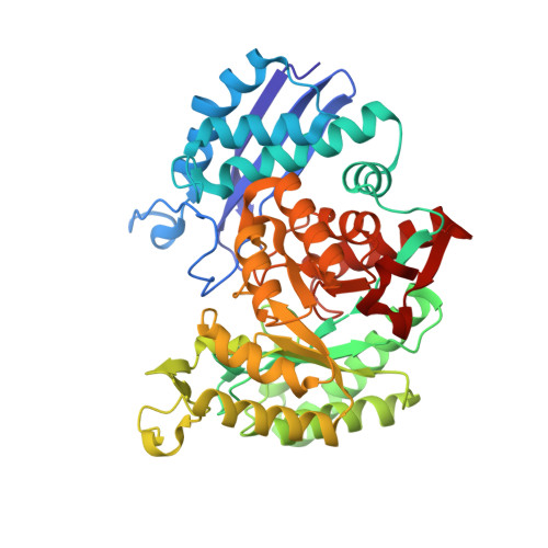
Crystallographic and kinetic methods have been used to characterize a site-specific variant of yeast enolase in which Ser 39 in the active-site flap has been changed to Ala. In the wild-type enzyme, the carbonyl and hydroxyl groups of Ser 39 chelate the second equivalent of divalent metal ion, effectively anchoring the flap over the fully liganded active site. With Mg(2+) as the activating cation, S39A enolase has <0.01% of wild-type activity as reported previously [J.M. Brewer, C.V. Glover, M.J. Holland, L. Lebioda, Biochim. Biophys. Acta 1383 (2) (1998) 351-355]. Measurements of (2)H kinetic isotope effects indicate that the proton abstraction from 2-phosphoglycerate (2-PGA) is significantly rate determining. Analysis of the isotope effects provides information on the relative rates of formation and breakdown of the enolate intermediate. Moreover, assays with different species of divalent metal ions reveal that with S39A enolase (unlike the case of wild-type enolase), more electrophilic metal ions promote higher activities. The kinetic results with the S39A variant support the notions that a rate-limiting product release lowers the activity of wild-type enolase with more electrophilic metal ions and that the metal ions are used to acidify the C2-proton of 2-PGA. The S39A enolase was co-crystallized with Mg(2+) and the inhibitor phosphonoacetohydroxamate (PhAH). The structure was solved and refined at a resolution of 2.1 A. The structure confirms the conjecture that the active-site flap is opened in the mutant protein. PhAH chelates to both Mg ions as in the corresponding structure of the wild-type complex. Positions of the side chains of catalytic groups, Lys 345 and Glu 211, and of "auxiliary" residues Glu 168 and Lys 396 are virtually unchanged relative to the complex with the wild-type protein. His 159, which hydrogen bonds to the phosphonate oxygens in the wild-type complex, is 5.7 A from the closest phosphonate oxygen, and the loop (154-166) containing His 159 is shifted away from the active center. A peripheral loop, Glu 251-Gly 275, also moves to open access to the active site.
Organizational Affiliation:
Department of Biochemistry, University of Wisconsin-Madison, Madison, WI 53705, USA.