1L8L

Molecular basis for the local confomational rearrangement of human phosphoserine phosphatase


Experimental Data Snapshot

  • Method: X-RAY DIFFRACTION
  • Resolution: 2.51 Å
  • R-Value Free: 0.284 
  • R-Value Work: 0.213 
  • R-Value Observed: 0.213 

wwPDB Validation   3D Report Full Report


This is version 1.3 of the entry. See complete history


Literature

Molecular basis for the local conformational rearrangement of human phosphoserine phosphatase.

Kim, H.Y.Heo, Y.S.Kim, J.H.Park, M.H.Moon, J.Kim, E.Kwon, D.Yoon, J.Shin, D.Jeong, E.J.Park, S.Y.Lee, T.G.Jeon, Y.H.Ro, S.Cho, J.M.Hwang, K.Y.

(2002) J Biol Chem 277: 46651-46658

  • DOI: https://doi.org/10.1074/jbc.M204866200
  • Primary Citation of Related Structures:  
    1L8L, 1L8O

  • PubMed Abstract: 

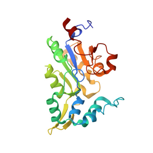
    Human phosphoserine phosphatase (HPSP) regulates the levels of glycine and d-serine, the putative co-agonists for the glycine site of the NMDA receptor in the brain. Here, we describe the first crystal structures of the HPSP in complexes with the competitive inhibitor 2-amino-3-phosphonopropionic acid (AP3) at 2.5 A, and the phosphate ion (Pi) and the product uncompetitive inhibitor l-serine (HPSP.l-Ser.Pi) at 2.8 A. The complex structures reveal that the open-closed environmental change of the active site, generated by local rearrangement of the alpha-helical bundle domain, is important to substrate recognition and hydrolysis. The maximal extent of this structural rearrangement is shown to be about 13 A at the L4 loop and about 25 degrees at the helix alpha3. Both the structural change and mutagenesis data suggest that Arg-65 and Glu-29 play an important role in the binding of the substrate. Interestingly, the AP3 binding mode turns out to be significantly different from that of the natural substrate, phospho-l-serine, and the HPSP.l-Ser.Pi structure provides a structural basis for the feedback control mechanism of serine. These analyses allow us to provide a clear model for the mechanism of HPSP and a framework for structure-based drug development.


  • Organizational Affiliation

    Divison of Drug Discovery, CrystalGenomics Incorporated, Daeduck Biocommunity, Jeonmin-dong, Yuseong-gu, Taejeon City, South Korea 305-600.


Macromolecules
Find similar proteins by:  (by identity cutoff)  |  3D Structure
Entity ID: 1
MoleculeChains Sequence LengthOrganismDetailsImage
L-3-phosphoserine phosphatase
A, B
225Homo sapiensMutation(s): 1 
EC: 3.1.3.3
UniProt & NIH Common Fund Data Resources
Find proteins for P78330 (Homo sapiens)
Explore P78330 
Go to UniProtKB:  P78330
PHAROS:  P78330
GTEx:  ENSG00000146733 
Entity Groups  
Sequence Clusters30% Identity50% Identity70% Identity90% Identity95% Identity100% Identity
UniProt GroupP78330
Sequence Annotations
Expand
  • Reference Sequence
Small Molecules
Ligands 1 Unique
IDChains Name / Formula / InChI Key2D Diagram3D Interactions
APO
Query on APO

Download Ideal Coordinates CCD File 
C [auth A],
D [auth B]
D-2-AMINO-3-PHOSPHONO-PROPIONIC ACID
C3 H8 N O5 P
LBTABPSJONFLPO-UWTATZPHSA-N
Experimental Data & Validation

Experimental Data

  • Method: X-RAY DIFFRACTION
  • Resolution: 2.51 Å
  • R-Value Free: 0.284 
  • R-Value Work: 0.213 
  • R-Value Observed: 0.213 
  • Space Group: I 4
Unit Cell:
Length ( Å )Angle ( ˚ )
a = 106.213α = 90
b = 106.213β = 90
c = 87.812γ = 90
Software Package:
Software NamePurpose
DENZOdata reduction
SCALEPACKdata scaling
CNSrefinement
CNSphasing

Structure Validation

View Full Validation Report


Entry History 

Deposition Data

Revision History  (Full details and data files)

  • Version 1.0: 2003-04-01
    Type: Initial release
  • Version 1.1: 2008-04-28
    Changes: Version format compliance
  • Version 1.2: 2011-07-13
    Changes: Version format compliance
  • Version 1.3: 2021-11-10
    Changes: Database references, Derived calculations