Biochemical characterization and structural analysis of a highly proficient cocaine esterase
Turner, J.M., Larsen, N.A., Basran, A., Barbas III, C.F., Bruce, N.C., Wilson, I.A., Lerner, R.A.(2002) Biochemistry 41: 12297-12307
- PubMed: 12369817
- DOI: https://doi.org/10.1021/bi026131p
- Primary Citation of Related Structures:
1L7Q, 1L7R - PubMed Abstract:
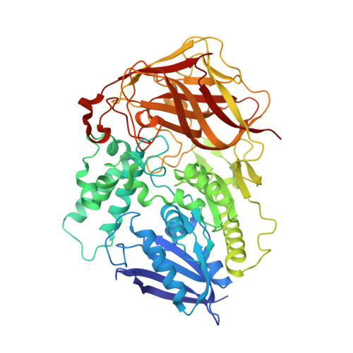
The bacterial cocaine esterase, cocE, hydrolyzes cocaine faster than any other reported cocaine esterase. Hydrolysis of the cocaine benzoyl ester follows Michaelis-Menten kinetics with k(cat) = 7.8 s(-1) and K(M) = 640 nM. A similar rate is observed for hydrolysis of cocaethylene, a more potent cocaine metabolite that has been observed in patients who concurrently abuse cocaine and alcohol. The high catalytic proficiency, lack of observable product inhibition, and ability to hydrolyze both cocaine and cocaethylene make cocE an attractive candidate for rapid cocaine detoxification in an emergency setting. Recently, we determined the crystal structure of this enzyme, and showed that it is a serine carboxylesterase, with a catalytic triad formed by S117, H287, and D259 within a hydrophobic active site, and an oxyanion hole formed by the backbone amide of Y118 and the Y44 hydroxyl. The only enzyme previously known to use a Tyr side chain to form the oxyanion hole is prolyl oligopeptidase, but the Y44F mutation of cocE has a more deleterious effect on the specificity rate constant (k(cat)/K(M)) than the analogous Y473F mutation of prolyl oligopeptidase. Kinetic studies on a series of cocE mutants both validate the proposed mechanism, and reveal the relative contributions of active site residues toward substrate recognition and catalysis. Inspired by the anionic binding pocket of the cocaine binding antibody GNC92H2, we found that a Q55E mutation within the active site of cocE results in a modest (2-fold) improvement in K(M), but a 14-fold loss of k(cat). The pH rate profile of cocE was fit to the ionization of two groups (pK(a1) = 7.7; pK(a2) = 10.4) that likely represent titration of H287 and Y44, respectively. We also describe the crystal structures of both S117A and Y44F mutants of cocE. Finally, urea denaturation studies of cocE by fluorescence and circular dichroism show two unfolding transitions (0.5-0.6 M and 3.2-3.7 M urea), with the first transition likely representing pertubation of the active site.
Organizational Affiliation:
Department of Molecular Biology, The Scripps Research Institute, 10550 North Torrey Pines Road, La Jolla, California 92037, USA.