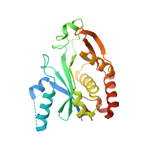
1KZF

Crystal Structure of the Acyl-homoserine Lactone Synthase, EsaI


Experimental Data Snapshot

  • Method: X-RAY DIFFRACTION
  • Resolution: 1.80 Å
  • R-Value Free: 0.244 
  • R-Value Work: 0.209 
  • R-Value Observed: 0.224 

wwPDB Validation   3D Report Full Report


This is version 1.3 of the entry. See complete history


Literature

Structural basis and specificity of acyl-homoserine lactone signal production in bacterial quorum sensing.

Watson, W.T.Minogue, T.D.Val, D.L.von Bodman, S.B.Churchill, M.E.

(2002) Mol Cell 9: 685-694

  • DOI: https://doi.org/10.1016/s1097-2765(02)00480-x
  • Primary Citation of Related Structures:  
    1K4J, 1KZF

  • PubMed Abstract: 

    Synthesis and detection of acyl-homoserine lactones (AHLs) enables many gram-negative bacteria to engage in quorum sensing, an intercellular signaling mechanism that activates differentiation to virulent and biofilm lifestyles. The AHL synthases catalyze acylation of S-adenosyl-L-methionine by acyl-acyl carrier protein and lactonization of the methionine moiety to give AHLs. The crystal structure of the AHL synthase, EsaI, determined at 1.8 A resolution, reveals a remarkable structural similarity to the N-acetyltransferases and defines a common phosphopantetheine binding fold as the catalytic core. Critical residues responsible for catalysis and acyl chain specificity have been identified from a modeled substrate complex and verified through functional analysis in vivo. A mechanism for the N-acylation of S-adenosyl-L-methionine by 3-oxo-hexanoyl-acyl carrier protein is proposed.


  • Organizational Affiliation

    Department of Pharmacology, The University of Colorado Health Sciences Center, 4200 E. Ninth Avenue, Denver, CO 80262, USA.


Macromolecules
Find similar proteins by:  (by identity cutoff)  |  3D Structure
Entity ID: 1
MoleculeChains Sequence LengthOrganismDetailsImage
acyl-homoserinelactone synthase EsaI230Pantoea stewartii subsp. stewartiiMutation(s): 0 
Gene Names: EsaI/EsaR gene cluster
UniProt
Find proteins for P54656 (Pantoea stewartii subsp. stewartii)
Explore P54656 
Go to UniProtKB:  P54656
Entity Groups  
Sequence Clusters30% Identity50% Identity70% Identity90% Identity95% Identity100% Identity
UniProt GroupP54656
Sequence Annotations
Expand
  • Reference Sequence
Experimental Data & Validation

Experimental Data

  • Method: X-RAY DIFFRACTION
  • Resolution: 1.80 Å
  • R-Value Free: 0.244 
  • R-Value Work: 0.209 
  • R-Value Observed: 0.224 
  • Space Group: P 43
Unit Cell:
Length ( Å )Angle ( ˚ )
a = 66.991α = 90
b = 66.991β = 90
c = 47.011γ = 90
Software Package:
Software NamePurpose
CNSrefinement
DENZOdata reduction
SCALEPACKdata scaling
CNSphasing

Structure Validation

View Full Validation Report


Entry History 

Deposition Data

Revision History  (Full details and data files)

  • Version 1.0: 2002-04-17
    Type: Initial release
  • Version 1.1: 2008-04-28
    Changes: Version format compliance
  • Version 1.2: 2011-07-13
    Changes: Version format compliance
  • Version 1.3: 2023-08-16
    Changes: Data collection, Database references, Refinement description