Crystal structure of thioltransferase at 2.2 A resolution.
Katti, S.K., Robbins, A.H., Yang, Y., Wells, W.W.(1995) Protein Sci 4: 1998-2005
- PubMed: 8535236
- DOI: https://doi.org/10.1002/pro.5560041005
- Primary Citation of Related Structures:
1KTE - PubMed Abstract:
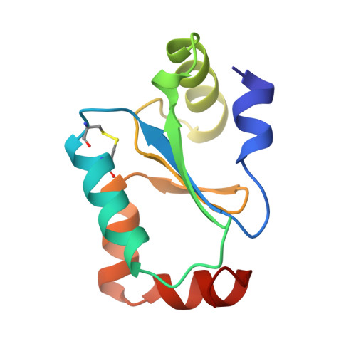
We report here the first three-dimensional structure of a mammalian thioltransferase as determined by single crystal X-ray crystallography at 2.2 A resolution. The protein is known for its thiol-redox properties and dehydroascorbate reductase activity. Recombinant pig liver thioltransferase expressed in Escherichia coli was crystallized in its oxidized form by vapor diffusion technique. The structure was determined by multiple isomorphous replacement method using four heavy-atom derivatives. The protein folds into an alpha/beta structure with a four-stranded mixed beta-sheet in the core, flanked on either side by helices. The fold is similar to that found in other thiol-redox proteins, viz. E. coli thioredoxin and bacteriophage T4 glutaredoxin, and thus seems to be conserved in these functionally related proteins. The active site disulfide (Cys 22-Cys 25) is located on a protrusion on the molecular surface. Cys 22, which is known to have an abnormally low pKa of 3.8, is accessible from the exterior of the molecule. Pro 70, which is in close proximity to the disulfide bridge, assumes a conserved cis-peptide configuration. Mutational data available on the protein are in agreement with the three-dimensional structure.
Organizational Affiliation:
Bayer Corporation, Pharmaceutical Division, West Haven, Connecticut 06516, USA.