1KQC

Structure of Nitroreductase from E. cloacae Complex with Inhibitor Acetate


Experimental Data Snapshot

  • Method: X-RAY DIFFRACTION
  • Resolution: 1.80 Å
  • R-Value Free: 0.219 
  • R-Value Work: 0.187 
  • R-Value Observed: 0.195 

wwPDB Validation   3D Report Full Report


Ligand Structure Quality Assessment 


This is version 1.4 of the entry. See complete history


Literature

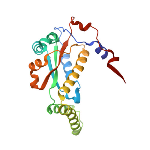
Structures of nitroreductase in three states: effects of inhibitor binding and reduction.

Haynes, C.A.Koder, R.L.Miller, A.F.Rodgers, D.W.

(2002) J Biol Chem 277: 11513-11520

  • DOI: https://doi.org/10.1074/jbc.M111334200
  • Primary Citation of Related Structures:  
    1KQB, 1KQC, 1KQD

  • PubMed Abstract: 

    The crystal structure of the nitroreductase enzyme from Enterobacter cloacae has been determined for the oxidized form in separate complexes with benzoate and acetate inhibitors and for the two-electron reduced form. Nitroreductase is a member of a group of enzymes that reduce a broad range of nitroaromatic compounds and has potential uses in chemotherapy and bioremediation. The monomers of the nitroreductase dimer adopt an alpha+beta fold and together bind two flavin mononucleotide prosthetic groups at the dimer interface. In the oxidized enzyme, the flavin ring system adopts a strongly bent (16 degrees ) conformation, and the bend increases (25 degrees ) in the reduced form of the enzyme, roughly the conformation predicted for reduced flavin free in solution. Because free oxidized flavin is planar, the induced bend in the oxidized enzyme may favor reduction, and it may also account for the characteristic inability of the enzyme to stabilize the one electron-reduced semiquinone flavin, which is also planar. Both inhibitors bind over the pyrimidine and central rings of the flavin in partially overlapping sites. Comparison of the two inhibitor complexes shows that a portion of helix H6 can flex to accommodate the differently sized inhibitors suggesting a mechanism for accommodating varied substrates.


  • Organizational Affiliation

    Department of Molecular and Cellular Biochemistry, The University of Kentucky, Lexington, Kentucky 40536, USA.


Macromolecules
Find similar proteins by:  (by identity cutoff)  |  3D Structure
Entity ID: 1
MoleculeChains Sequence LengthOrganismDetailsImage
OXYGEN-INSENSITIVE NAD(P)H NITROREDUCTASE
A, B, C, D
217Enterobacter cloacaeMutation(s): 0 
EC: 1.6.6
UniProt
Find proteins for Q01234 (Enterobacter cloacae)
Explore Q01234 
Go to UniProtKB:  Q01234
Entity Groups  
Sequence Clusters30% Identity50% Identity70% Identity90% Identity95% Identity100% Identity
UniProt GroupQ01234
Sequence Annotations
Expand
  • Reference Sequence
Experimental Data & Validation

Experimental Data

  • Method: X-RAY DIFFRACTION
  • Resolution: 1.80 Å
  • R-Value Free: 0.219 
  • R-Value Work: 0.187 
  • R-Value Observed: 0.195 
  • Space Group: P 1 21 1
Unit Cell:
Length ( Å )Angle ( ˚ )
a = 52.785α = 90
b = 79.606β = 93.63
c = 97.138γ = 90
Software Package:
Software NamePurpose
CNSrefinement
SCALEPACKdata scaling
CNSphasing

Structure Validation

View Full Validation Report


Ligand Structure Quality Assessment 


Entry History 

Deposition Data

Revision History  (Full details and data files)

  • Version 1.0: 2002-02-13
    Type: Initial release
  • Version 1.1: 2008-04-27
    Changes: Version format compliance
  • Version 1.2: 2011-07-13
    Changes: Version format compliance
  • Version 1.3: 2017-10-11
    Changes: Refinement description
  • Version 1.4: 2024-02-14
    Changes: Data collection, Database references, Derived calculations