High-resolution crystal structures of the lectin-like xylan binding domain from Streptomyces lividans xylanase 10A with bound substrates reveal a novel mode of xylan binding.
Notenboom, V., Boraston, A.B., Williams, S.J., Kilburn, D.G., Rose, D.R.(2002) Biochemistry 41: 4246-4254
- PubMed: 11914070
- DOI: https://doi.org/10.1021/bi015865j
- Primary Citation of Related Structures:
1KNL, 1KNM, 1MC9 - PubMed Abstract:
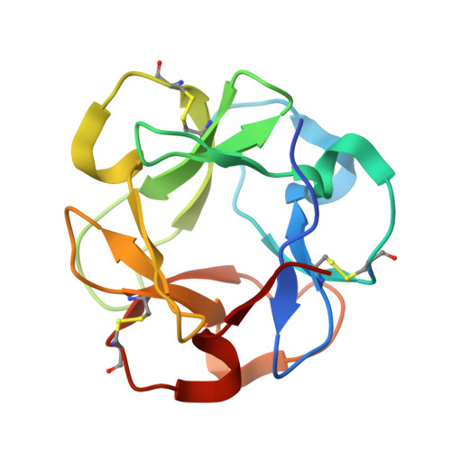
Carbohydrate-binding module (CBM) family 13 includes the "R-type" or "ricin superfamily" beta-trefoil lectins. The C-terminal CBM, CBM13, of xylanase 10A from Streptomyces lividans is a family 13 CBM that is not only structurally similar to the "R-type" lectins but also somewhat functionally similar. The primary function of CBM13 is to bind the polysaccharide xylan, but it retains the ability of the R-type lectins to bind small sugars such as lactose and galactose. The association of CBM13 with xylan appears to involve cooperative and additive participation of three binding pockets in each of the three trefoil domains of CBM13, suggesting a novel mechanism of CBM-xylan interaction. Thus, the interaction of CBM13 with sugars displays considerable plasticity for which we provide a structural rationale. The high-resolution crystal structure of CBM13 was determined by multiple anomalous dispersion from a complex of CBM13 with a brominated ligand. Crystal structures of CBM13 in complex with lactose and xylopentaose revealed two distinct mechanisms of ligand binding. CBM13 has retained its specificity for lactose via Ricin-like binding in all of the three classic trefoil binding pockets. However, CBM13 has the ability to bind either the nonreducing galactosyl moiety or the reducing glucosyl moiety of lactose. The mode of xylopentaose binding suggests adaptive mutations in the trefoil sugar binding scaffold to accommodate internal binding on helical polymers of xylose.
Organizational Affiliation:
Protein Engineering Networks of Centres of Excellence and Department of Microbiology and Immunology and Biotechnology Laboratory, University of British Columbia, Vancouver, BC, Canada. valerie@nki.nl