1KIJ

Crystal structure of the 43K ATPase domain of Thermus thermophilus gyrase B in complex with novobiocin


Experimental Data Snapshot

  • Method: X-RAY DIFFRACTION
  • Resolution: 2.30 Å
  • R-Value Free: 0.266 
  • R-Value Work: 0.206 
  • R-Value Observed: 0.206 

wwPDB Validation   3D Report Full Report


Ligand Structure Quality Assessment 


This is version 1.4 of the entry. See complete history


Literature

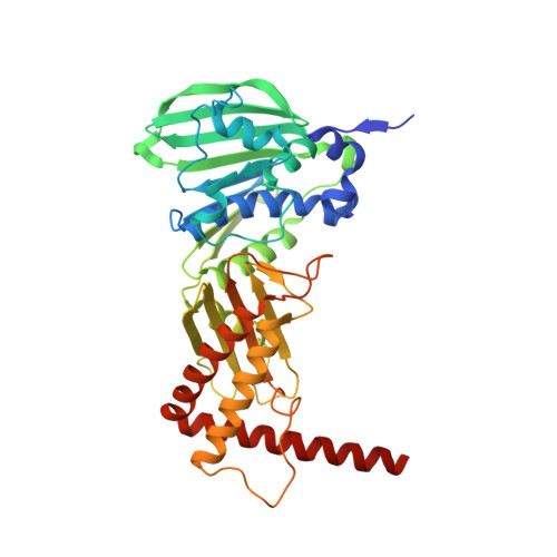
An open conformation of the Thermus thermophilus gyrase B ATP-binding domain.

Lamour, V.Hoermann, L.Jeltsch, J.M.Oudet, P.Moras, D.

(2002) J Biol Chem 277: 18947-18953

  • DOI: https://doi.org/10.1074/jbc.M111740200
  • Primary Citation of Related Structures:  
    1KIJ

  • PubMed Abstract: 

    DNA gyrase forms an A(2)B(2) tetramer involved in DNA replication, repair, recombination, and transcription in which the B subunit catalyzes ATP hydrolysis. The Thermus thermophilus and Escherichia coli gyrases are homologues and present the same catalytic activity. When compared with that of the E. coli 43K-5'-adenylyl-beta,gamma-imidodiphosphate complex, the crystal structure of Gyrase B 43K ATPase domain in complex with novobiocin, one of the most potent inhibitors of gyrase shows large conformational changes of the subdomains within the dimer. The stabilization of loop 98-118 closing the active site through dimeric contacts and interaction with domain 2 allows to observe novobiocin-protein interactions that could not be seen in the 24K-inhibitor complexes. Furthermore, this loop adopts a position which defines an "open" conformation of the active site in absence of ATP, in contrast with the "closed" conformation adopted upon ATP binding. All together, these results indicate how the subdomains may propagate conformational changes from the active site and provide crucial information for the design of more specific inhibitors.


  • Organizational Affiliation

    Institut de Génétique et de Biologie Moléculaire, CNRS/INSERM/ULP, BP163, 1 rue Laurent Fries, 67404 Illkirch Cedex, France.


Macromolecules
Find similar proteins by:  (by identity cutoff)  |  3D Structure
Entity ID: 1
MoleculeChains Sequence LengthOrganismDetailsImage
DNA GYRASE SUBUNIT B
A, B
390Thermus thermophilusMutation(s): 0 
EC: 5.99.1.3
UniProt
Find proteins for Q5SHZ4 (Thermus thermophilus (strain ATCC 27634 / DSM 579 / HB8))
Explore Q5SHZ4 
Go to UniProtKB:  Q5SHZ4
Entity Groups  
Sequence Clusters30% Identity50% Identity70% Identity90% Identity95% Identity100% Identity
UniProt GroupQ5SHZ4
Sequence Annotations
Expand
  • Reference Sequence
Experimental Data & Validation

Experimental Data

  • Method: X-RAY DIFFRACTION
  • Resolution: 2.30 Å
  • R-Value Free: 0.266 
  • R-Value Work: 0.206 
  • R-Value Observed: 0.206 
  • Space Group: P 1 21 1
Unit Cell:
Length ( Å )Angle ( ˚ )
a = 44.88α = 90
b = 125.55β = 96.36
c = 79.83γ = 90
Software Package:
Software NamePurpose
AMoREphasing
CNSrefinement
DENZOdata reduction
SCALEPACKdata scaling

Structure Validation

View Full Validation Report


Ligand Structure Quality Assessment 


Entry History 

Deposition Data

Revision History  (Full details and data files)

  • Version 1.0: 2002-06-03
    Type: Initial release
  • Version 1.1: 2008-04-27
    Changes: Version format compliance
  • Version 1.2: 2011-07-13
    Changes: Non-polymer description, Version format compliance
  • Version 1.3: 2016-06-01
    Changes: Source and taxonomy
  • Version 1.4: 2024-02-14
    Changes: Data collection, Database references, Derived calculations