1KGT

Crystal Structure of Tetrahydrodipicolinate N-Succinyltransferase in Complex with Pimelate and Succinyl-CoA


Experimental Data Snapshot

  • Method: X-RAY DIFFRACTION
  • Resolution: 2.30 Å
  • R-Value Free: 0.261 
  • R-Value Work: 0.168 
  • R-Value Observed: 0.171 

wwPDB Validation   3D Report Full Report


This is version 1.3 of the entry. See complete history


Literature

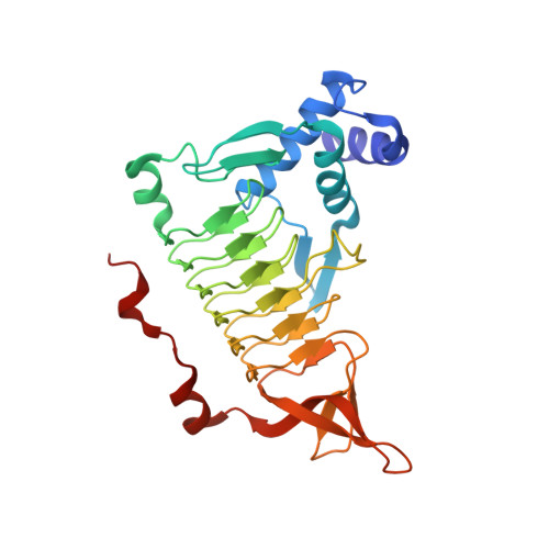
Acyl group specificity at the active site of tetrahydridipicolinate N-succinyltransferase.

Beaman, T.W.Vogel, K.W.Drueckhammer, D.G.Blanchard, J.S.Roderick, S.L.

(2002) Protein Sci 11: 974-979

  • DOI: https://doi.org/10.1110/ps.4310102
  • Primary Citation of Related Structures:  
    1KGQ, 1KGT

  • PubMed Abstract: 

    Tetrahydrodipicolinate N-succinyltransferase (DapD) catalyzes the succinyl-CoA-dependent acylation of L-2-amino-6-oxopimelate to 2-N-succinyl-6-oxopimelate as part of the succinylase branch of the meso-diaminopimelate/lysine biosynthetic pathway of bacteria, blue-green algae, and plants. This pathway provides meso-diaminopimelate as a building block for cell wall peptidoglycan in most bacteria, and is regarded as a target pathway for antibacterial agents. We have solved the X-ray crystal structures of DapD in ternary complexes with pimelate/succinyl-CoA and L-2-aminopimelate with the nonreactive cofactor analog, succinamide-CoA. These structures define the binding conformation of the cofactor succinyl group and its interactions with the enzyme and place its thioester carbonyl carbon in close proximity to the nucleophilic 2-amino group of the acceptor, in support of a direct attack ternary complex mechanism. The acyl group specificity differences between homologous tetrahydrodipicolinate N-acetyl- and N-succinyltransferases can be rationalized with reference to at least three amino acids that interact with or give accessible active site volume to the cofactor succinyl group. These residues account at least in part for the substrate specificity that commits metabolic intermediates to either the succinylase or acetylase branches of the meso-diaminopimelate/lysine biosynthetic pathway.


  • Organizational Affiliation

    Department of Biochemistry, Albert Einstein College of Medicine, Bronx, New York 10461, USA.


Macromolecules
Find similar proteins by:  (by identity cutoff)  |  3D Structure
Entity ID: 1
MoleculeChains Sequence LengthOrganismDetailsImage
2,3,4,5-TETRAHYDROPYRIDINE-2-CARBOXYLATE N-SUCCINYLTRANSFERASE274Mycobacterium tuberculosis variant bovisMutation(s): 0 
Gene Names: dapD
EC: 2.3.1.117
UniProt
Find proteins for P56220 (Unknown prokaryotic organism)
Explore P56220 
Go to UniProtKB:  P56220
Entity Groups  
Sequence Clusters30% Identity50% Identity70% Identity90% Identity95% Identity100% Identity
UniProt GroupP56220
Sequence Annotations
Expand
  • Reference Sequence
Small Molecules
Ligands 2 Unique
IDChains Name / Formula / InChI Key2D Diagram3D Interactions
SCA
Query on SCA

Download Ideal Coordinates CCD File 
C [auth A]SUCCINYL-COENZYME A
C25 H40 N7 O19 P3 S
VNOYUJKHFWYWIR-ITIYDSSPSA-N
PML
Query on PML

Download Ideal Coordinates CCD File 
B [auth A]PIMELIC ACID
C7 H12 O4
WLJVNTCWHIRURA-UHFFFAOYSA-N
Experimental Data & Validation

Experimental Data

  • Method: X-RAY DIFFRACTION
  • Resolution: 2.30 Å
  • R-Value Free: 0.261 
  • R-Value Work: 0.168 
  • R-Value Observed: 0.171 
  • Space Group: H 3
Unit Cell:
Length ( Å )Angle ( ˚ )
a = 95.98α = 90
b = 95.98β = 90
c = 73.44γ = 120
Software Package:
Software NamePurpose
TNTrefinement
X-GENdata reduction
XDSdata scaling
TNTphasing

Structure Validation

View Full Validation Report


Entry History 

Deposition Data

Revision History  (Full details and data files)

  • Version 1.0: 2002-04-03
    Type: Initial release
  • Version 1.1: 2008-04-27
    Changes: Version format compliance
  • Version 1.2: 2011-07-13
    Changes: Derived calculations, Version format compliance
  • Version 1.3: 2024-02-14
    Changes: Data collection, Database references, Derived calculations, Refinement description