Crystallographic and thiol-reactivity studies on the complex of pig muscle phosphoglycerate kinase with ATP analogues: correlation between nucleotide binding mode and helix flexibility.
Kovari, Z., Flachner, B., Naray-Szabo, G., Vas, M.(2002) Biochemistry 41: 8796-8806
- PubMed: 12102622
- DOI: https://doi.org/10.1021/bi020210j
- Primary Citation of Related Structures:
1KF0 - PubMed Abstract:
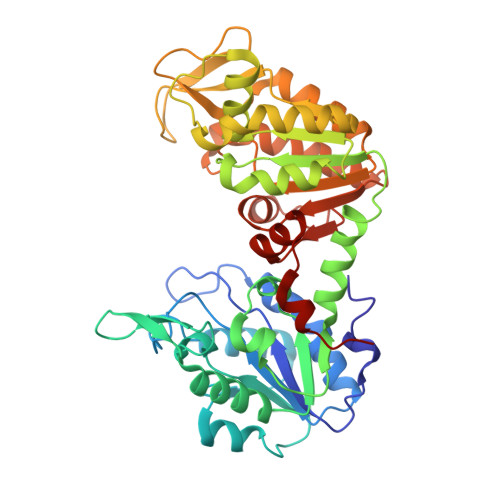
Crystal structure of the ternary complex of pig muscle phosphoglycerate kinase (PGK) with the substrate 3-phosphoglycerate (3-PG) and the Mg(2+) complex of beta,gamma-methylene-adenosine-5'-triphosphate (AMP-PCP), a nonreactive analogue of the nucleotide substrate, MgATP, has been determined by X-ray diffraction at 2.5 A resolution. The overall structure of the protein exhibits an open conformation, similar to that of the previously determined ternary complex of the pig muscle enzyme with beta,gamma-imido-adenosine-5'-triphosphate (AMP-PNP) in place of AMP-PCP (May, Vas, Harlos, and Blake (1996) Proteins 24, 292-303). The orientation and details of interactions of the nucleotide phosphates, however, show marked differences. The beta-phosphate is linked to the conserved Asp 218, i.e., to the N-terminus of helix 8, through the Mg(2+) ion; the previously observed interactions of the metal complex of AMP-PNP or ADP with the conserved Asn 336 and the N-terminus of helix 13 are completely absent. These structural differences are maintained themselves in solution studies. Inhibition and binding experiments show a slightly weaker interaction of PGK with MgAMP-PCP than with MgAMP-PNP: at pH 7.5, the K(d) values are 1.07 +/- 0.18 and 0.41 +/- 0.08 mM, respectively. The difference is further enhanced by 3-PG: the K(d) values are 2.80 +/- 0.66 and 0.68 +/- 0.11 mM, respectively. Thus, the previously observed weakening effect of 3-PG on nucleotide binding (Merli, Szilágyi, Flachner, Rossi, and Vas (2002) Biochemistry 41, 111-119) is more pronounced with MgAMP-PCP. The discordance between substrate analogues also shows up in thiol reactivity studies. In their binary complexes, both ATP analogues protect the fast-reacting thiols of PGK in helix 13 against modification to similar extent. In their ternary complexes, however, which also contain bound 3-PG, the protective effect of MgAMP-PCP, but not of MgAMP-PNP, is largely abolished. This indicates a much smaller effect of MgAMP-PCP on the conformation of helix 13, which is in good correlation with its altered mode of phosphate binding and the ensuing increase in the flexibility of helix 13, as shown by elevated crystallographic B-factors. The possible existence of alternative site(s) for binding of the nucleotide phosphates may have functional relevance.
Organizational Affiliation:
Eötvös Loránd University, Department of Theoretical Chemistry, H-1518, Budapest 112, P.O. Box 32, Hungary.