1KAW

STRUCTURE OF SINGLE STRANDED DNA BINDING PROTEIN (SSB)


Experimental Data Snapshot

  • Method: X-RAY DIFFRACTION
  • Resolution: 2.90 Å
  • R-Value Free: 0.289 
  • R-Value Work: 0.221 
  • R-Value Observed: 0.221 

wwPDB Validation   3D Report Full Report


This is version 1.3 of the entry. See complete history


Literature

Crystal structure of the homo-tetrameric DNA binding domain of Escherichia coli single-stranded DNA-binding protein determined by multiwavelength x-ray diffraction on the selenomethionyl protein at 2.9-A resolution.

Raghunathan, S.Ricard, C.S.Lohman, T.M.Waksman, G.

(1997) Proc Natl Acad Sci U S A 94: 6652-6657

  • DOI: https://doi.org/10.1073/pnas.94.13.6652
  • Primary Citation of Related Structures:  
    1KAW

  • PubMed Abstract: 

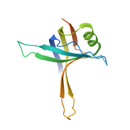
    The crystal structure of the tetrameric DNA-binding domain of the single-stranded DNA binding protein from Escherichia coli was determined at a resolution of 2.9 A using multiwavelength anomalous dispersion. Each monomer in the tetramer is topologically similar to an oligomer-binding fold. Two monomers each contribute three beta-strands to a single six-stranded beta-sheet to form a dimer. Two dimer-dimer interfaces are observed within the crystal. One of these stabilizes the tetramer in solution. The other interface promotes a superhelical structure within the crystal that may reflect tetramer-tetramer interactions involved in the positive cooperative binding of the single-stranded DNA-binding protein to single-stranded DNA.


  • Organizational Affiliation

    Department of Biochemistry and Molecular Biophysics, Washington University School of Medicine, St. Louis, MO 63110, USA.


Macromolecules
Find similar proteins by:  (by identity cutoff)  |  3D Structure
Entity ID: 1
MoleculeChains Sequence LengthOrganismDetailsImage
SINGLE-STRANDED DNA BINDING PROTEIN
A, B, C, D
135Escherichia coliMutation(s): 0 
UniProt
Find proteins for P0AGE0 (Escherichia coli (strain K12))
Explore P0AGE0 
Go to UniProtKB:  P0AGE0
Entity Groups  
Sequence Clusters30% Identity50% Identity70% Identity90% Identity95% Identity100% Identity
UniProt GroupP0AGE0
Sequence Annotations
Expand
  • Reference Sequence
Experimental Data & Validation

Experimental Data

  • Method: X-RAY DIFFRACTION
  • Resolution: 2.90 Å
  • R-Value Free: 0.289 
  • R-Value Work: 0.221 
  • R-Value Observed: 0.221 
  • Space Group: C 1 2 1
Unit Cell:
Length ( Å )Angle ( ˚ )
a = 58.08α = 90
b = 105.85β = 99
c = 90.24γ = 90
Software Package:
Software NamePurpose
X-PLORmodel building
X-PLORrefinement
DENZOdata reduction
SCALEPACKdata scaling
X-PLORphasing

Structure Validation

View Full Validation Report


Entry History 

Deposition Data

Revision History  (Full details and data files)

  • Version 1.0: 1997-12-31
    Type: Initial release
  • Version 1.1: 2008-03-24
    Changes: Version format compliance
  • Version 1.2: 2011-07-13
    Changes: Version format compliance
  • Version 1.3: 2024-02-07
    Changes: Data collection, Database references, Other, Structure summary