1KAG

Crystal Structure of the Escherichia coli Shikimate Kinase I (AroK)


Experimental Data Snapshot

  • Method: X-RAY DIFFRACTION
  • Resolution: 2.05 Å
  • R-Value Free: 0.271 
  • R-Value Work: 0.217 
  • R-Value Observed: 0.217 

wwPDB Validation   3D Report Full Report


This is version 1.4 of the entry. See complete history


Literature

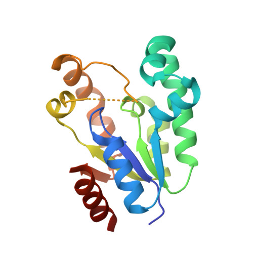
Crystal structure of the Escherichia coli shikimate kinase I (AroK) that confers sensitivity to mecillinam.

Romanowski, M.J.Burley, S.K.

(2002) Proteins 47: 558-562


Macromolecules
Find similar proteins by:  (by identity cutoff)  |  3D Structure
Entity ID: 1
MoleculeChains Sequence LengthOrganismDetailsImage
Shikimate kinase I
A, B
173Escherichia coliMutation(s): 0 
Gene Names: AROK
EC: 2.7.1.71
UniProt
Find proteins for P0A6D7 (Escherichia coli (strain K12))
Explore P0A6D7 
Go to UniProtKB:  P0A6D7
Entity Groups  
Sequence Clusters30% Identity50% Identity70% Identity90% Identity95% Identity100% Identity
UniProt GroupP0A6D7
Sequence Annotations
Expand
  • Reference Sequence
Experimental Data & Validation

Experimental Data

  • Method: X-RAY DIFFRACTION
  • Resolution: 2.05 Å
  • R-Value Free: 0.271 
  • R-Value Work: 0.217 
  • R-Value Observed: 0.217 
  • Space Group: P 1 21 1
Unit Cell:
Length ( Å )Angle ( ˚ )
a = 35.726α = 90
b = 63.417β = 91.27
c = 67.941γ = 90
Software Package:
Software NamePurpose
SnBphasing
MLPHAREphasing
DMmodel building
CNSrefinement
MARMADdata reduction
DENZOdata reduction
SCALEPACKdata scaling
DMphasing

Structure Validation

View Full Validation Report


Entry History 

Deposition Data

Revision History  (Full details and data files)

  • Version 1.0: 2002-05-08
    Type: Initial release
  • Version 1.1: 2008-04-27
    Changes: Version format compliance
  • Version 1.2: 2011-07-13
    Changes: Version format compliance
  • Version 1.3: 2021-02-03
    Changes: Database references, Refinement description, Structure summary
  • Version 1.4: 2024-02-07
    Changes: Data collection, Database references