1K9O

CRYSTAL STRUCTURE OF MICHAELIS SERPIN-TRYPSIN COMPLEX


Experimental Data Snapshot

  • Method: X-RAY DIFFRACTION
  • Resolution: 2.30 Å
  • R-Value Free: 0.231 
  • R-Value Work: 0.158 

wwPDB Validation   3D Report Full Report


This is version 1.4 of the entry. See complete history


Literature

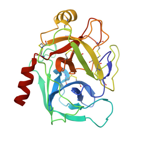
The structure of a Michaelis serpin-protease complex.

Ye, S.Cech, A.L.Belmares, R.Bergstrom, R.C.Tong, Y.Corey, D.R.Kanost, M.R.Goldsmith, E.J.

(2001) Nat Struct Biol 8: 979-983

  • DOI: https://doi.org/10.1038/nsb1101-979
  • Primary Citation of Related Structures:  
    1K9O

  • PubMed Abstract: 

    Serine protease inhibitors (serpins) regulate the activities of circulating proteases. Serpins inhibit proteases by acylating the serine hydroxyl at their active sites. Before deacylation and complete proteolysis of the serpin can occur, massive conformational changes are triggered in the serpin while maintaining the covalent linkage between the protease and serpin. Here we report the structure of a serpin-trypsin Michaelis complex, which we visualized by using the S195A trypsin mutant to prevent covalent complex formation. This encounter complex reveals a more extensive interaction surface than that present in small inhibitor-protease complexes and is a template for modeling other serpin-protease pairs. Mutations of several serpin residues at the interface reduced the inhibitory activity of the serpin. The serine residue C-terminal to the scissile peptide bond is found in a closer than usual interaction with His 57 at the active site of trypsin.


  • Organizational Affiliation

    Department of Biochemistry, The University of Texas Southwestern Medical Center at Dallas, 5323 Harry Hines Boulevard, Dallas, Texas 75390, USA.


Macromolecules
Find similar proteins by:  (by identity cutoff)  |  3D Structure
Entity ID: 1
MoleculeChains Sequence LengthOrganismDetailsImage
ALASERPINA [auth I]378Manduca sextaMutation(s): 1 
UniProt
Find proteins for P14754 (Manduca sexta)
Explore P14754 
Go to UniProtKB:  P14754
Entity Groups  
Sequence Clusters30% Identity50% Identity70% Identity90% Identity95% Identity100% Identity
UniProt GroupP14754
Sequence Annotations
Expand
  • Reference Sequence
Find similar proteins by:  (by identity cutoff)  |  3D Structure
Entity ID: 2
MoleculeChains Sequence LengthOrganismDetailsImage
TRYPSIN II ANIONICB [auth E]223Rattus norvegicusMutation(s): 1 
EC: 3.4.21.4
UniProt
Find proteins for P00763 (Rattus norvegicus)
Explore P00763 
Go to UniProtKB:  P00763
Entity Groups  
Sequence Clusters30% Identity50% Identity70% Identity90% Identity95% Identity100% Identity
UniProt GroupP00763
Sequence Annotations
Expand
  • Reference Sequence
Experimental Data & Validation

Experimental Data

  • Method: X-RAY DIFFRACTION
  • Resolution: 2.30 Å
  • R-Value Free: 0.231 
  • R-Value Work: 0.158 
  • Space Group: P 31 2 1
Unit Cell:
Length ( Å )Angle ( ˚ )
a = 112.572α = 90
b = 112.572β = 90
c = 95.955γ = 120
Software Package:
Software NamePurpose
DENZOdata reduction
SCALEPACKdata scaling
AMoREphasing
CNSrefinement

Structure Validation

View Full Validation Report


Entry History 

Deposition Data

Revision History  (Full details and data files)

  • Version 1.0: 2001-11-21
    Type: Initial release
  • Version 1.1: 2008-04-27
    Changes: Version format compliance
  • Version 1.2: 2011-07-13
    Changes: Version format compliance
  • Version 1.3: 2021-10-27
    Changes: Database references
  • Version 1.4: 2023-08-16
    Changes: Data collection, Refinement description