1K96

CRYSTAL STRUCTURE OF CALCIUM BOUND HUMAN S100A6


Experimental Data Snapshot

  • Method: X-RAY DIFFRACTION
  • Resolution: 1.44 Å
  • R-Value Free: 0.226 
  • R-Value Work: 0.214 
  • R-Value Observed: 0.214 

wwPDB Validation   3D Report Full Report


This is version 1.3 of the entry. See complete history


Literature

Crystal structures of S100A6 in the Ca(2+)-free and Ca(2+)-bound states: the calcium sensor mechanism of S100 proteins revealed at atomic resolution.

Otterbein, L.R.Kordowska, J.Witte-Hoffmann, C.Wang, C.L.Dominguez, R.

(2002) Structure 10: 557-567

  • DOI: https://doi.org/10.1016/s0969-2126(02)00740-2
  • Primary Citation of Related Structures:  
    1K8U, 1K96, 1K9K, 1K9P

  • PubMed Abstract: 

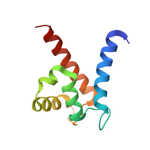
    S100A6 is a member of the S100 family of Ca(2+) binding proteins, which have come to play an important role in the diagnosis of cancer due to their overexpression in various tumor cells. We have determined the crystal structures of human S100A6 in the Ca(2+)-free and Ca(2+)-bound states to resolutions of 1.15 A and 1.44 A, respectively. Ca(2+) binding is responsible for a dramatic change in the global shape and charge distribution of the S100A6 dimer, leading to the exposure of two symmetrically positioned target binding sites. The results are consistent with S100A6, and most likely other S100 proteins, functioning as Ca(2+) sensors in a way analogous to the prototypical sensors calmodulin and troponin C. The structures have important implications for our understanding of target binding and cooperativity of Ca(2+) binding in the S100 family.


  • Organizational Affiliation

    Boston Biomedical Research Institute, 64 Grove Street, Watertown, MA 02472, USA.


Macromolecules
Find similar proteins by:  (by identity cutoff)  |  3D Structure
Entity ID: 1
MoleculeChains Sequence LengthOrganismDetailsImage
S100A690Homo sapiensMutation(s): 0 
UniProt & NIH Common Fund Data Resources
Find proteins for P06703 (Homo sapiens)
Explore P06703 
Go to UniProtKB:  P06703
PHAROS:  P06703
GTEx:  ENSG00000197956 
Entity Groups  
Sequence Clusters30% Identity50% Identity70% Identity90% Identity95% Identity100% Identity
UniProt GroupP06703
Sequence Annotations
Expand
  • Reference Sequence
Small Molecules
Ligands 2 Unique
IDChains Name / Formula / InChI Key2D Diagram3D Interactions
BME
Query on BME

Download Ideal Coordinates CCD File 
D [auth A]BETA-MERCAPTOETHANOL
C2 H6 O S
DGVVWUTYPXICAM-UHFFFAOYSA-N
CA
Query on CA

Download Ideal Coordinates CCD File 
B [auth A],
C [auth A]
CALCIUM ION
Ca
BHPQYMZQTOCNFJ-UHFFFAOYSA-N
Experimental Data & Validation

Experimental Data

  • Method: X-RAY DIFFRACTION
  • Resolution: 1.44 Å
  • R-Value Free: 0.226 
  • R-Value Work: 0.214 
  • R-Value Observed: 0.214 
  • Space Group: C 1 2 1
Unit Cell:
Length ( Å )Angle ( ˚ )
a = 45.18α = 90
b = 39.23β = 110.44
c = 48.11γ = 90
Software Package:
Software NamePurpose
AMoREphasing
REFMACrefinement
HKL-2000data reduction
HKL-2000data scaling

Structure Validation

View Full Validation Report


Entry History 

Deposition Data

Revision History  (Full details and data files)

  • Version 1.0: 2002-04-10
    Type: Initial release
  • Version 1.1: 2008-04-27
    Changes: Version format compliance
  • Version 1.2: 2011-07-13
    Changes: Derived calculations, Version format compliance
  • Version 1.3: 2023-08-16
    Changes: Data collection, Database references, Derived calculations, Refinement description