1K6W

The Structure of Escherichia coli Cytosine Deaminase


Experimental Data Snapshot

  • Method: X-RAY DIFFRACTION
  • Resolution: 1.75 Å
  • R-Value Free: 0.172 
  • R-Value Work: 0.155 
  • R-Value Observed: 0.157 

wwPDB Validation   3D Report Full Report


This is version 1.4 of the entry. See complete history


Literature

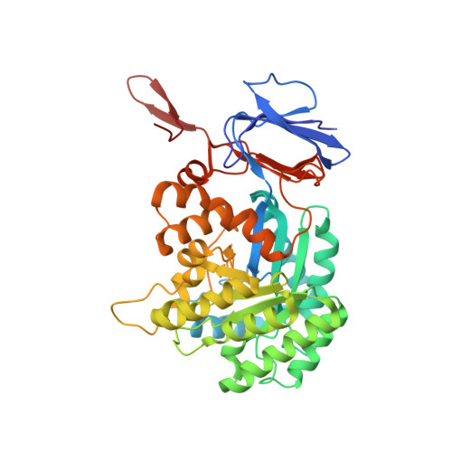
The structure of Escherichia coli cytosine deaminase.

Ireton, G.C.McDermott, G.Black, M.E.Stoddard, B.L.

(2002) J Mol Biol 315: 687-697

  • DOI: https://doi.org/10.1006/jmbi.2001.5277
  • Primary Citation of Related Structures:  
    1K6W, 1K70

  • PubMed Abstract: 

    Cytosine deaminase (CD) catalyzes the deamination of cytosine, producing uracil. This enzyme is present in prokaryotes and fungi (but not multicellular eukaryotes) and is an important member of the pyrimidine salvage pathway in those organisms. The same enzyme also catalyzes the conversion of 5-fluorocytosine to 5-fluorouracil; this activity allows the formation of a cytotoxic chemotherapeutic agent from a non-cytotoxic precursor. The enzyme is of widespread interest both for antimicrobial drug design and for gene therapy applications against tumors. The structure of Escherichia coli CD has been determined in the presence and absence of a bound mechanism-based inhibitor. The enzyme forms an (alphabeta)(8) barrel structure with structural similarity to adenosine deaminase, a relationship that is undetectable at the sequence level, and no similarity to bacterial cytidine deaminase. The enzyme is packed into a hexameric assembly stabilized by a unique domain-swapping interaction between enzyme subunits. The active site is located in the mouth of the enzyme barrel and contains a bound iron ion that coordinates a hydroxyl nucleophile. Substrate binding involves a significant conformational change that sequesters the reaction complex from solvent.


  • Organizational Affiliation

    Fred Hutchinson Cancer Research Center and the Graduate Program in Molecular and Cell Biology, University of Washington, 1100 Fairview Ave. N. A3-023, Seattle, WA 98109, USA.


Macromolecules
Find similar proteins by:  (by identity cutoff)  |  3D Structure
Entity ID: 1
MoleculeChains Sequence LengthOrganismDetailsImage
Cytosine Deaminase426Escherichia coliMutation(s): 0 
EC: 3.5.4.1
UniProt
Find proteins for P25524 (Escherichia coli (strain K12))
Explore P25524 
Go to UniProtKB:  P25524
Entity Groups  
Sequence Clusters30% Identity50% Identity70% Identity90% Identity95% Identity100% Identity
UniProt GroupP25524
Sequence Annotations
Expand
  • Reference Sequence
Small Molecules
Ligands 1 Unique
IDChains Name / Formula / InChI Key2D Diagram3D Interactions
FE
Query on FE

Download Ideal Coordinates CCD File 
B [auth A]FE (III) ION
Fe
VTLYFUHAOXGGBS-UHFFFAOYSA-N
Experimental Data & Validation

Experimental Data

  • Method: X-RAY DIFFRACTION
  • Resolution: 1.75 Å
  • R-Value Free: 0.172 
  • R-Value Work: 0.155 
  • R-Value Observed: 0.157 
  • Space Group: H 3 2
Unit Cell:
Length ( Å )Angle ( ˚ )
a = 109.15α = 90
b = 109.15β = 90
c = 240.05γ = 120
Software Package:
Software NamePurpose
CNSrefinement
DENZOdata reduction
SCALEPACKdata scaling
CNSphasing

Structure Validation

View Full Validation Report


Entry History 

Deposition Data

Revision History  (Full details and data files)

  • Version 1.0: 2002-02-06
    Type: Initial release
  • Version 1.1: 2008-04-27
    Changes: Version format compliance
  • Version 1.2: 2011-07-13
    Changes: Derived calculations, Version format compliance
  • Version 1.3: 2011-11-16
    Changes: Atomic model
  • Version 1.4: 2024-02-07
    Changes: Data collection, Database references, Derived calculations