1K4W

X-ray structure of the orphan nuclear receptor ROR beta ligand-binding domain in the active conformation


Experimental Data Snapshot

  • Method: X-RAY DIFFRACTION
  • Resolution: 1.90 Å
  • R-Value Free: 0.249 
  • R-Value Work: 0.224 

wwPDB Validation   3D Report Full Report


Ligand Structure Quality Assessment 


This is version 1.4 of the entry. See complete history


Literature

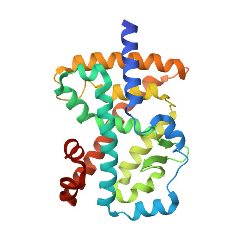
X-ray structure of the orphan nuclear receptor RORbeta ligand-binding domain in the active conformation.

Stehlin, C.Wurtz, J.M.Steinmetz, A.Greiner, E.Schule, R.Moras, D.Renaud, J.P.

(2001) EMBO J 20: 5822-5831

  • DOI: https://doi.org/10.1093/emboj/20.21.5822
  • Primary Citation of Related Structures:  
    1K4W

  • PubMed Abstract: 

    The retinoic acid-related orphan receptor beta (RORbeta) exhibits a highly restricted neuronal-specific expression pattern in brain, retina and pineal gland. So far, neither a natural RORbeta target gene nor a functional ligand have been identified, and the physiological role of the receptor is not well understood. We present the crystal structure of the ligand-binding domain (LBD) of RORbeta containing a bound stearate ligand and complexed with a coactivator peptide. In the crystal, the monomeric LBD adopts the canonical agonist-bound form. The fatty acid ligand-coactivator peptide combined action stabilizes the transcriptionally active conformation. The large ligand-binding pocket is strictly hydrophobic on the AF-2 side and more polar on the beta-sheet side where the carboxylate group of the ligand binds. Site-directed mutagenesis experiments validate the significance of the present structure. Homology modeling of the other isotypes will help to design isotype-selective agonists and antagonists that can be used to characterize the physiological functions of RORs. In addition, our crystallization strategy can be extended to other orphan nuclear receptors, providing a powerful tool to delineate their functions.


  • Organizational Affiliation

    Laboratoire de Biologie et Génomique Structurales (CNRS Unité Propre de Recherche 9004), Institut de Génétique et de Biologie Moléculaire et Cellulaire (CNRS/INSERM/Université Louis Pasteur), 1 rue Laurent Fries, BP 163, 67404 Illkirch, France.


Macromolecules
Find similar proteins by:  (by identity cutoff)  |  3D Structure
Entity ID: 1
MoleculeChains Sequence LengthOrganismDetailsImage
Nuclear receptor ROR-beta252Rattus norvegicusMutation(s): 0 
Gene Names: NR1F2
UniProt
Find proteins for P45446 (Rattus norvegicus)
Explore P45446 
Go to UniProtKB:  P45446
Entity Groups  
Sequence Clusters30% Identity50% Identity70% Identity90% Identity95% Identity100% Identity
UniProt GroupP45446
Sequence Annotations
Expand
  • Reference Sequence

Find similar proteins by:  Sequence   |   3D Structure  

Entity ID: 2
MoleculeChains Sequence LengthOrganismDetailsImage
steroid receptor coactivator-115N/AMutation(s): 0 
UniProt & NIH Common Fund Data Resources
Find proteins for Q15788 (Homo sapiens)
Explore Q15788 
Go to UniProtKB:  Q15788
PHAROS:  Q15788
GTEx:  ENSG00000084676 
Entity Groups  
Sequence Clusters30% Identity50% Identity70% Identity90% Identity95% Identity100% Identity
UniProt GroupQ15788
Sequence Annotations
Expand
  • Reference Sequence
Small Molecules
Ligands 1 Unique
IDChains Name / Formula / InChI Key2D Diagram3D Interactions
STE
Query on STE

Download Ideal Coordinates CCD File 
C [auth A]STEARIC ACID
C18 H36 O2
QIQXTHQIDYTFRH-UHFFFAOYSA-N
Experimental Data & Validation

Experimental Data

  • Method: X-RAY DIFFRACTION
  • Resolution: 1.90 Å
  • R-Value Free: 0.249 
  • R-Value Work: 0.224 
  • Space Group: P 21 21 21
Unit Cell:
Length ( Å )Angle ( ˚ )
a = 52.302α = 90
b = 58.489β = 90
c = 106.036γ = 90
Software Package:
Software NamePurpose
AMoREphasing
CNSrefinement
DENZOdata reduction
SCALEPACKdata scaling

Structure Validation

View Full Validation Report


Ligand Structure Quality Assessment 


Entry History 

Deposition Data

Revision History  (Full details and data files)

  • Version 1.0: 2002-04-09
    Type: Initial release
  • Version 1.1: 2008-04-27
    Changes: Version format compliance
  • Version 1.2: 2011-07-13
    Changes: Version format compliance
  • Version 1.3: 2021-07-21
    Changes: Derived calculations, Refinement description
  • Version 1.4: 2023-08-16
    Changes: Data collection, Database references, Refinement description