1K3C

Phosphoenolpyruvate carboxykinase in complex with ADP, AlF3 and Pyruvate


Experimental Data Snapshot

  • Method: X-RAY DIFFRACTION
  • Resolution: 2.00 Å
  • R-Value Free: 0.285 
  • R-Value Work: 0.223 
  • R-Value Observed: 0.225 

wwPDB Validation   3D Report Full Report


Ligand Structure Quality Assessment 


This is version 2.0 of the entry. See complete history


Literature

The phosphoryl-transfer mechanism of Escherichia coli phosphoenolpyruvate carboxykinase from the use of AlF(3).

Sudom, A.M.Prasad, L.Goldie, H.Delbaere, L.T.

(2001) J Mol Biol 314: 83-92

  • DOI: https://doi.org/10.1006/jmbi.2001.5120
  • Primary Citation of Related Structures:  
    1K3C, 1K3D

  • PubMed Abstract: 

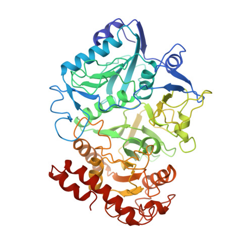
    The mechanism of reversible transfer of the gamma-phosphate group of ATP by Escherichia coli phosphoenolpyruvate carboxykinase (PCK) on to its substrate is of great interest. It is known that metallofluorides are accurate analogs of the transition state in the context of kinase mechanisms. Therefore, two complexes of PCK, one with AlF(3), Mg(2+) and ADP (complex I), the other with AlF(3), Mg(2+), ADP and pyruvate (complex II) were crystallized. The X-ray crystal structures of these two complexes were determined at 2.0 A resolution. The Al atom has trigonal bipyramidal geometry that mimics the transition state of phosphoryl transfer. The Al atom is at a distance of 2.8 A and 2.9 A from an oxygen atom of the beta-phosphoryl group of ADP in complex I and II, respectively. A water molecule in complex I and an oxygen atom of the pyruvate in complex II are located along the axis of the trigonal bipyramid on the side opposite to the beta-phosphoryl oxygen with respect to the equatorial plane, suggesting that the complexes are close mimics of the transition state. Along with the presence of positively charged species around the AlF(3) moiety, these results indicate that phosphoryl transfer occurs via a direct displacement mechanism with associative qualities.


  • Organizational Affiliation

    Department of Biochemistry, University of Saskatchewan, Saskatoon, Canada.


Macromolecules
Find similar proteins by:  (by identity cutoff)  |  3D Structure
Entity ID: 1
MoleculeChains Sequence LengthOrganismDetailsImage
Phosphoenolpyruvate carboxykinase540Escherichia coliMutation(s): 0 
Gene Names: PCKA
EC: 4.1.1.49
UniProt
Find proteins for P22259 (Escherichia coli (strain K12))
Explore P22259 
Go to UniProtKB:  P22259
Entity Groups  
Sequence Clusters30% Identity50% Identity70% Identity90% Identity95% Identity100% Identity
UniProt GroupP22259
Sequence Annotations
Expand
  • Reference Sequence
Experimental Data & Validation

Experimental Data

  • Method: X-RAY DIFFRACTION
  • Resolution: 2.00 Å
  • R-Value Free: 0.285 
  • R-Value Work: 0.223 
  • R-Value Observed: 0.225 
  • Space Group: C 1 2 1
Unit Cell:
Length ( Å )Angle ( ˚ )
a = 124.738α = 90
b = 94.211β = 96.05
c = 46.336γ = 90
Software Package:
Software NamePurpose
AMoREphasing
X-PLORrefinement
DENZOdata reduction
SCALEPACKdata scaling

Structure Validation

View Full Validation Report


Ligand Structure Quality Assessment 


Entry History 

Deposition Data

Revision History  (Full details and data files)

  • Version 1.0: 2001-12-19
    Type: Initial release
  • Version 1.1: 2008-04-27
    Changes: Version format compliance
  • Version 1.2: 2011-07-13
    Changes: Version format compliance
  • Version 1.3: 2023-08-16
    Changes: Data collection, Database references, Derived calculations, Refinement description
  • Version 2.0: 2023-11-15
    Changes: Atomic model, Data collection