The crystallographic structure of brome mosaic virus.
Lucas, R.W., Larson, S.B., McPherson, A.(2002) J Mol Biol 317: 95-108
- PubMed: 11916381
- DOI: https://doi.org/10.1006/jmbi.2001.5389
- Primary Citation of Related Structures:
1JS9 - PubMed Abstract:
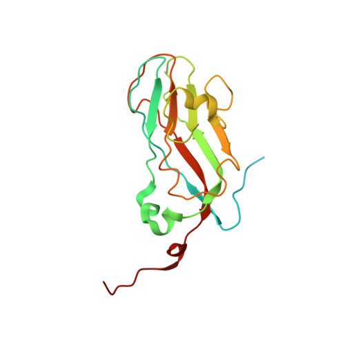
The structure of brome mosaic virus (BMV), the type member of the bromoviridae family, has been determined from a single rhombohedral crystal by X-ray diffraction, and refined to an R value of 0.237 for data in the range 3.4-40.0 A. The structure, which represents the native, compact form at pH 5.2 in the presence of 0.1 M Mg(2+), was solved by molecular replacement using the model of cowpea chlorotic mottle virus (CCMV), which BMV closely resembles. The BMV model contains amino acid residues 41-189 for the pentameric capsid A subunits, and residues 25-189 and 1-189 for the B and C subunits, respectively, which compose the hexameric capsomeres. In the model there are two Mg ions and one molecule of polyethylene glycol (PEG). The first 25 amino acid residues of the C subunit are modeled as polyalanine. The coat protein has the canonical "jellyroll" beta-barrel topology with extended amino-terminal polypeptides as seen in other icosahedral plant viruses. Mass spectrometry shows that in native BMV virions, a significant fraction of the amino-terminal peptides are apparently cleaved. No recognizable nucleic acid residue is visible in the electron density maps except at low resolution where it appears to exhibit a layered arrangement in the virion interior. It is juxtaposed closely with the interior surface of the capsid but does not interpenetrate. The protein subunits forming hexameric capsomeres, and particularly dimers, appear to interact extensively, but the subunits otherwise contact one another sparsely about the 5-fold and quasi 3-fold axes. Thus, the virion appears to be an assembly of loosely associated hexameric capsomeres, which may be the basis for the swelling and dissociation that occurs at neutral pH and elevated salt concentration. A Mg ion is observed to lie exactly on the quasi-3-fold axis and is closely coordinated by side-chains of three quasi-symmetry-related residues glutamates 84, with possible participation of side-chains from threonines 145, and asparagines 148. A presumptive Mg(2+) is also present on the 5-fold axis where there is a concentration of negatively charged side-chains, but the precise coordination is unclear. In both cases these cations appear to be essential for maintenance of virion stability. Density that is contiguous with the viral interior is present on the 3-fold axis at the center of the hexameric capsomere, where there is a pore of about 6 A diameter. The density cannot be attributed to cations and it was modeled as a PEG molecule.
Organizational Affiliation:
University of California-Irvine, 560 Steinhaus Hall, Irvine, CA 92697-3900, USA.