1JRO

Crystal Structure of Xanthine Dehydrogenase from Rhodobacter capsulatus


Experimental Data Snapshot

  • Method: X-RAY DIFFRACTION
  • Resolution: 2.70 Å
  • R-Value Free: 0.252 
  • R-Value Work: 0.213 
  • R-Value Observed: 0.215 

wwPDB Validation   3D Report Full Report


This is version 1.4 of the entry. See complete history


Literature

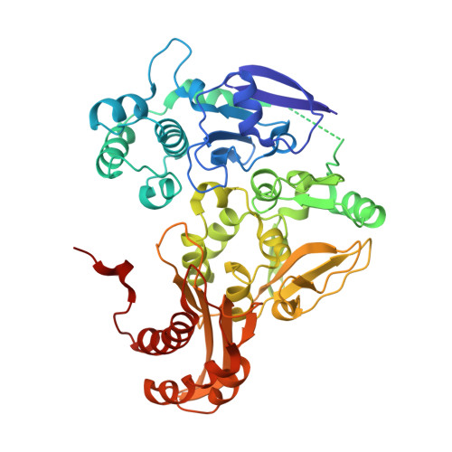
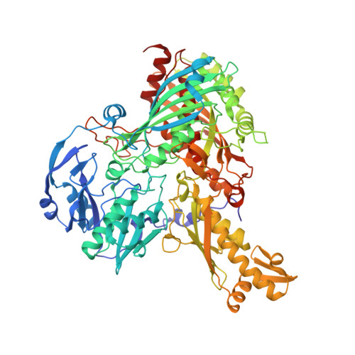
Crystal structures of the active and alloxanthine-inhibited forms of xanthine dehydrogenase from Rhodobacter capsulatus

Truglio, J.J.Theis, K.Leimkuhler, S.Rappa, R.Rajagopalan, K.V.Kisker, C.

(2002) Structure 10: 115-125

  • DOI: https://doi.org/10.1016/s0969-2126(01)00697-9
  • Primary Citation of Related Structures:  
    1JRO, 1JRP

  • PubMed Abstract: 

    Xanthine dehydrogenase (XDH), a complex molybdo/iron-sulfur/flavoprotein, catalyzes the oxidation of hypoxanthine to xanthine followed by oxidation of xanthine to uric acid with concomitant reduction of NAD+. The 2.7 A resolution structure of Rhodobacter capsulatus XDH reveals that the bacterial and bovine XDH have highly similar folds despite differences in subunit composition. The NAD+ binding pocket of the bacterial XDH resembles that of the dehydrogenase form of the bovine enzyme rather than that of the oxidase form, which reduces O(2) instead of NAD+. The drug allopurinol is used to treat XDH-catalyzed uric acid build-up occurring in gout or during cancer chemotherapy. As a hypoxanthine analog, it is oxidized to alloxanthine, which cannot be further oxidized but acts as a tight binding inhibitor of XDH. The 3.0 A resolution structure of the XDH-alloxanthine complex shows direct coordination of alloxanthine to the molybdenum via a nitrogen atom. These results provide a starting point for the rational design of new XDH inhibitors.


  • Organizational Affiliation

    Department of Pharmacological Sciences, Center for Structural Biology, State University of New York at Stony Brook, Stony Brook, NY 11794, USA.


Macromolecules
Find similar proteins by:  (by identity cutoff)  |  3D Structure
Entity ID: 1
MoleculeChains Sequence LengthOrganismDetailsImage
xanthine dehydrogenase, chain A
A, C, E, G
462Rhodobacter capsulatusMutation(s): 0 
EC: 1.1.1.204
UniProt
Find proteins for O54050 (Rhodobacter capsulatus)
Explore O54050 
Go to UniProtKB:  O54050
Entity Groups  
Sequence Clusters30% Identity50% Identity70% Identity90% Identity95% Identity100% Identity
UniProt GroupO54050
Sequence Annotations
Expand
  • Reference Sequence
Find similar proteins by:  (by identity cutoff)  |  3D Structure
Entity ID: 2
MoleculeChains Sequence LengthOrganismDetailsImage
xanthine dehydrogenase, chain B
B, D, F, H
777Rhodobacter capsulatusMutation(s): 0 
EC: 1.1.1.204
UniProt
Find proteins for O54051 (Rhodobacter capsulatus)
Explore O54051 
Go to UniProtKB:  O54051
Entity Groups  
Sequence Clusters30% Identity50% Identity70% Identity90% Identity95% Identity100% Identity
UniProt GroupO54051
Sequence Annotations
Expand
  • Reference Sequence
Small Molecules
Ligands 5 Unique
IDChains Name / Formula / InChI Key2D Diagram3D Interactions
FAD
Query on FAD

Download Ideal Coordinates CCD File 
CA [auth G],
K [auth A],
Q [auth C],
W [auth E]
FLAVIN-ADENINE DINUCLEOTIDE
C27 H33 N9 O15 P2
VWWQXMAJTJZDQX-UYBVJOGSSA-N
MTE
Query on MTE

Download Ideal Coordinates CCD File 
EA [auth H],
M [auth B],
S [auth D],
Y [auth F]
PHOSPHONIC ACIDMONO-(2-AMINO-5,6-DIMERCAPTO-4-OXO-3,7,8A,9,10,10A-HEXAHYDRO-4H-8-OXA-1,3,9,10-TETRAAZA-ANTHRACEN-7-YLMETHYL)ESTER
C10 H14 N5 O6 P S2
HPEUEJRPDGMIMY-IFQPEPLCSA-N
FES
Query on FES

Download Ideal Coordinates CCD File 
AA [auth G]
BA [auth G]
I [auth A]
J [auth A]
O [auth C]
AA [auth G],
BA [auth G],
I [auth A],
J [auth A],
O [auth C],
P [auth C],
U [auth E],
V [auth E]
FE2/S2 (INORGANIC) CLUSTER
Fe2 S2
NIXDOXVAJZFRNF-UHFFFAOYSA-N
MOS
Query on MOS

Download Ideal Coordinates CCD File 
FA [auth H],
N [auth B],
T [auth D],
Z [auth F]
DIOXOTHIOMOLYBDENUM(VI) ION
H Mo O2 S
BDSRWPHSAKXXRG-UHFFFAOYSA-M
CA
Query on CA

Download Ideal Coordinates CCD File 
DA [auth H],
L [auth B],
R [auth D],
X [auth F]
CALCIUM ION
Ca
BHPQYMZQTOCNFJ-UHFFFAOYSA-N
Experimental Data & Validation

Experimental Data

  • Method: X-RAY DIFFRACTION
  • Resolution: 2.70 Å
  • R-Value Free: 0.252 
  • R-Value Work: 0.213 
  • R-Value Observed: 0.215 
  • Space Group: P 1
Unit Cell:
Length ( Å )Angle ( ˚ )
a = 92.875α = 109.53
b = 141.053β = 105.83
c = 158.113γ = 101.33
Software Package:
Software NamePurpose
DENZOdata reduction
SCALEPACKdata scaling
AMoREphasing
REFMACrefinement

Structure Validation

View Full Validation Report


Entry History 

Deposition Data

Revision History  (Full details and data files)

  • Version 1.0: 2002-01-11
    Type: Initial release
  • Version 1.1: 2008-04-27
    Changes: Version format compliance
  • Version 1.2: 2011-07-13
    Changes: Version format compliance
  • Version 1.3: 2015-04-22
    Changes: Non-polymer description
  • Version 1.4: 2023-08-16
    Changes: Data collection, Database references, Derived calculations, Refinement description