1JR8

Crystal Structure of Erv2p


Experimental Data Snapshot

  • Method: X-RAY DIFFRACTION
  • Resolution: 1.50 Å
  • R-Value Free: 0.233 
  • R-Value Work: 0.208 

wwPDB Validation   3D Report Full Report


Ligand Structure Quality Assessment 


This is version 1.2 of the entry. See complete history


Literature

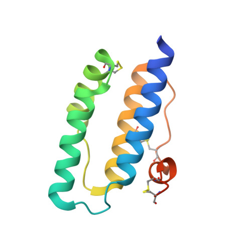
A new FAD-binding fold and intersubunit disulfide shuttle in the thiol oxidase Erv2p.

Gross, E.Sevier, C.S.Vala, A.Kaiser, C.A.Fass, D.

(2002) Nat Struct Biol 9: 61-67

  • DOI: https://doi.org/10.1038/nsb740
  • Primary Citation of Related Structures:  
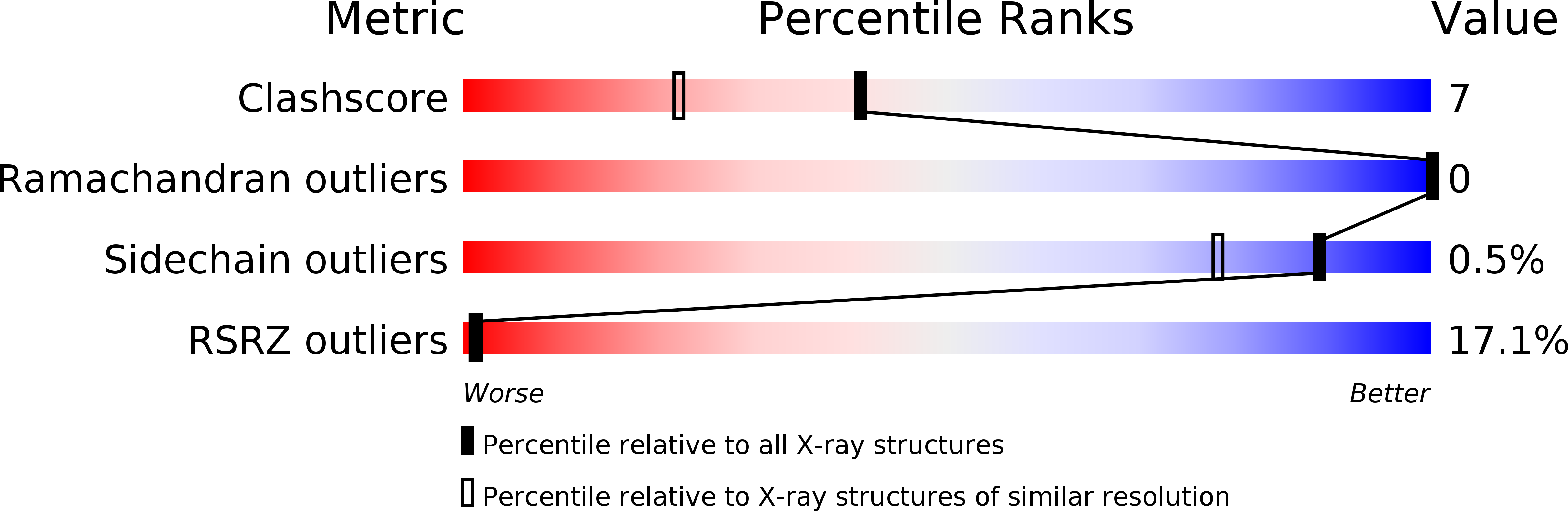
    1JR8, 1JRA

  • PubMed Abstract: 

    Erv2p is an FAD-dependent sulfhydryl oxidase that can promote disulfide bond formation during protein biosynthesis in the yeast endoplasmic reticulum. The structure of Erv2p, determined by X-ray crystallography to 1.5 A resolution, reveals a helix-rich dimer with no global resemblance to other known FAD-binding proteins or thiol oxidoreductases. Two pairs of cysteine residues are required for Erv2p activity. The first (Cys-Gly-Glu-Cys) is adjacent to the isoalloxazine ring of the FAD. The second (Cys-Gly-Cys) is part of a flexible C-terminal segment that can swing into the vicinity of the first cysteine pair in the opposite subunit of the dimer and may shuttle electrons between substrate protein dithiols and the FAD-proximal disulfide.


  • Organizational Affiliation

    Department of Structural Biology, Weizmann Institute of Science, Rehovot 76100, Israel.


Macromolecules
Find similar proteins by:  (by identity cutoff)  |  3D Structure
Entity ID: 1
MoleculeChains Sequence LengthOrganismDetailsImage
Erv2 PROTEIN, mitochondrial
A, B
117Saccharomyces cerevisiaeMutation(s): 0 
Gene Names: ERV2
UniProt
Find proteins for Q12284 (Saccharomyces cerevisiae (strain ATCC 204508 / S288c))
Explore Q12284 
Go to UniProtKB:  Q12284
Entity Groups  
Sequence Clusters30% Identity50% Identity70% Identity90% Identity95% Identity100% Identity
UniProt GroupQ12284
Sequence Annotations
Expand
  • Reference Sequence
Small Molecules
Ligands 1 Unique
IDChains Name / Formula / InChI Key2D Diagram3D Interactions
FAD
Query on FAD

Download Ideal Coordinates CCD File 
C [auth A],
D [auth B]
FLAVIN-ADENINE DINUCLEOTIDE
C27 H33 N9 O15 P2
VWWQXMAJTJZDQX-UYBVJOGSSA-N
Experimental Data & Validation

Experimental Data

  • Method: X-RAY DIFFRACTION
  • Resolution: 1.50 Å
  • R-Value Free: 0.233 
  • R-Value Work: 0.208 
  • Space Group: P 1 21 1
Unit Cell:
Length ( Å )Angle ( ˚ )
a = 47.64α = 90
b = 45.15β = 100.15
c = 53.84γ = 90
Software Package:
Software NamePurpose
DENZOdata reduction
SCALEPACKdata scaling
SOLVEphasing
CNSrefinement

Structure Validation

View Full Validation Report


Ligand Structure Quality Assessment 


Entry History 

Deposition Data

Revision History  (Full details and data files)

  • Version 1.0: 2001-12-28
    Type: Initial release
  • Version 1.1: 2008-04-27
    Changes: Version format compliance
  • Version 1.2: 2011-07-13
    Changes: Version format compliance