1JOU

Crystal Structure of Native S195A Thrombin with an Unoccupied Active Site


Experimental Data Snapshot

  • Method: X-RAY DIFFRACTION
  • Resolution: 1.80 Å
  • R-Value Free: 0.245 
  • R-Value Work: 0.222 

wwPDB Validation   3D Report Full Report


Ligand Structure Quality Assessment 


This is version 2.2 of the entry. See complete history


Literature

The molecular basis of thrombin allostery revealed by a 1.8A structure of the slow form

Huntington, J.A.Esmon, C.T.

(2003) Structure 11: 469-479

  • DOI: https://doi.org/10.1016/s0969-2126(03)00049-2
  • Primary Citation of Related Structures:  
    1JOU

  • PubMed Abstract: 

    Thrombin participates in its own positive and negative feedback loops, and its allosteric state helps determine the hemostatic balance. Here we present the 1.8 A crystallographic structure of S195A thrombin in two conformational states: active site occupied and active site free. The active site-occupied form shows how thrombin can accommodate substrates, such as protein C. The active site-free form is in a previously unobserved closed conformation of thrombin, which satisfies all the conditions of the so-called "slow" form. A mechanism of allostery is revealed, which relies on the concerted movement of the disulphide bond between Cys168 and 182 and aromatic residues Phe227, Trp215, and Trp60d. These residues constitute an allosteric switch, which is flipped directly through sodium binding, resulting in the fast form with an open active site.


  • Organizational Affiliation

    Department of Haematology, University of Cambridge, Cambridge Institute for Medical Research, Wellcome Trust/MRC Building, Hills Road, CB2 2XY, Cambridge, United Kingdom. jah52@cam.ac.uk


Macromolecules
Find similar proteins by:  (by identity cutoff)  |  3D Structure
Entity ID: 1
MoleculeChains Sequence LengthOrganismDetailsImage
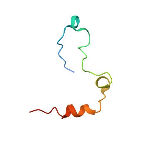
Thrombin, light chain
A, C, E
49Homo sapiensMutation(s): 0 
UniProt & NIH Common Fund Data Resources
Find proteins for P00734 (Homo sapiens)
Explore P00734 
Go to UniProtKB:  P00734
PHAROS:  P00734
GTEx:  ENSG00000180210 
Entity Groups  
Sequence Clusters30% Identity50% Identity70% Identity90% Identity95% Identity100% Identity
UniProt GroupP00734
Sequence Annotations
Expand
  • Reference Sequence
Find similar proteins by:  (by identity cutoff)  |  3D Structure
Entity ID: 2
MoleculeChains Sequence LengthOrganismDetailsImage
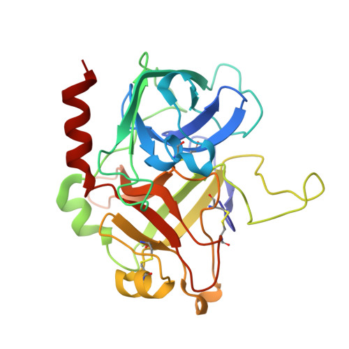
Thrombin, heavy chain
B, D, F
259Homo sapiensMutation(s): 1 
UniProt & NIH Common Fund Data Resources
Find proteins for P00734 (Homo sapiens)
Explore P00734 
Go to UniProtKB:  P00734
PHAROS:  P00734
GTEx:  ENSG00000180210 
Entity Groups  
Sequence Clusters30% Identity50% Identity70% Identity90% Identity95% Identity100% Identity
UniProt GroupP00734
Sequence Annotations
Expand
  • Reference Sequence
Small Molecules
Ligands 4 Unique
IDChains Name / Formula / InChI Key2D Diagram3D Interactions
NAG
Query on NAG

Download Ideal Coordinates CCD File 
G [auth B],
Q [auth F]
2-acetamido-2-deoxy-beta-D-glucopyranose
C8 H15 N O6
OVRNDRQMDRJTHS-FMDGEEDCSA-N
GOL
Query on GOL

Download Ideal Coordinates CCD File 
I [auth B],
J [auth B],
K [auth B],
L [auth B]
GLYCEROL
C3 H8 O3
PEDCQBHIVMGVHV-UHFFFAOYSA-N
ACY
Query on ACY

Download Ideal Coordinates CCD File 
M [auth B],
N [auth B],
P [auth D],
S [auth F],
T [auth F]
ACETIC ACID
C2 H4 O2
QTBSBXVTEAMEQO-UHFFFAOYSA-N
NA
Query on NA

Download Ideal Coordinates CCD File 
H [auth B],
O [auth D],
R [auth F]
SODIUM ION
Na
FKNQFGJONOIPTF-UHFFFAOYSA-N
Experimental Data & Validation

Experimental Data

  • Method: X-RAY DIFFRACTION
  • Resolution: 1.80 Å
  • R-Value Free: 0.245 
  • R-Value Work: 0.222 
  • Space Group: P 1 21 1
Unit Cell:
Length ( Å )Angle ( ˚ )
a = 71.23α = 90
b = 76.58β = 106.64
c = 97.32γ = 90
Software Package:
Software NamePurpose
MOSFLMdata reduction
SCALAdata scaling
MOLREPphasing
CNSrefinement
CCP4data scaling

Structure Validation

View Full Validation Report


Ligand Structure Quality Assessment 


Entry History 

Deposition Data

Revision History  (Full details and data files)

  • Version 1.0: 2001-08-10
    Type: Initial release
  • Version 1.1: 2008-04-27
    Changes: Version format compliance
  • Version 1.2: 2011-07-13
    Changes: Non-polymer description, Version format compliance
  • Version 2.0: 2020-07-29
    Type: Remediation
    Reason: Carbohydrate remediation
    Changes: Advisory, Atomic model, Data collection, Derived calculations, Non-polymer description, Structure summary
  • Version 2.1: 2021-10-27
    Changes: Advisory, Database references, Structure summary
  • Version 2.2: 2023-08-16
    Changes: Data collection, Refinement description