1JL0

Structure of a Human S-Adenosylmethionine Decarboxylase Self-processing Ester Intermediate and Mechanism of Putrescine Stimulation of Processing as Revealed by the H243A Mutant


Experimental Data Snapshot

  • Method: X-RAY DIFFRACTION
  • Resolution: 1.50 Å
  • R-Value Free: 0.237 
  • R-Value Work: 0.215 
  • R-Value Observed: 0.221 

wwPDB Validation   3D Report Full Report


This is version 1.5 of the entry. See complete history


Literature

Structure of a human S-adenosylmethionine decarboxylase self-processing ester intermediate and mechanism of putrescine stimulation of processing as revealed by the H243A mutant.

Ekstrom, J.L.Tolbert, W.D.Xiong, H.Pegg, A.E.Ealick, S.E.

(2001) Biochemistry 40: 9495-9504

  • DOI: https://doi.org/10.1021/bi010736o
  • Primary Citation of Related Structures:  
    1JL0

  • PubMed Abstract: 

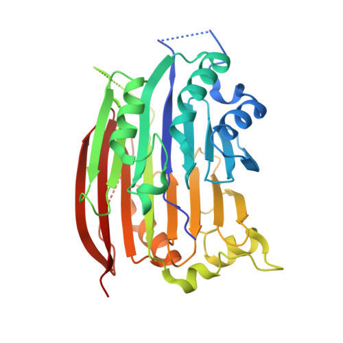
    S-Adenosylmethionine decarboxylase (AdoMetDC) is synthesized as a proenzyme that cleaves itself in a putrescine-stimulated reaction via an N-->O acyl shift and beta-elimination to produce an active enzyme with a catalytically essential pyruvoyl residue at the new N-terminus. N-->O acyl shifts initiate the self-processing of other proteins such as inteins and amidohydrolases, but their mechanisms in such proteins are not well understood. We have solved the crystal structure of the H243A mutant of AdoMetDC to 1.5 A resolution. The mutant protein is trapped in the ester form, providing clear evidence for the structure of the ester intermediate in the processing of pyruvoyl enzymes. In addition, a putrescine molecule is bound in a charged region within the beta-sandwich, and cross-links the two beta-sheets through hydrogen bonds to several acidic residues and ordered water molecules. The high-resolution structure provides insight into the mechanism for the self-processing reaction and provides evidence for the mechanism for simulation of the self-processing reaction by putrescine. Studies of the effects of putrescine or 4-aminobutanol on the processing of mutant AdoMetDC proenzymes are consistent with a model in which a single activator molecule interacts with buried Asp174, Glu178, and Glu256, leading to an alteration in the position of Glu11, resulting in stimulation of self-processing.


  • Organizational Affiliation

    Department of Chemistry and Chemical Biology, Cornell University, Ithaca, New York 14853-1301, USA.


Macromolecules
Find similar proteins by:  (by identity cutoff)  |  3D Structure
Entity ID: 1
MoleculeChains Sequence LengthOrganismDetailsImage
S-ADENOSYLMETHIONINE DECARBOXYLASE PROENZYME
A, B
334Homo sapiensMutation(s): 1 
EC: 4.1.1.50
UniProt & NIH Common Fund Data Resources
Find proteins for P17707 (Homo sapiens)
Explore P17707 
Go to UniProtKB:  P17707
PHAROS:  P17707
GTEx:  ENSG00000123505 
Entity Groups  
Sequence Clusters30% Identity50% Identity70% Identity90% Identity95% Identity100% Identity
UniProt GroupP17707
Sequence Annotations
Expand
  • Reference Sequence
Experimental Data & Validation

Experimental Data

  • Method: X-RAY DIFFRACTION
  • Resolution: 1.50 Å
  • R-Value Free: 0.237 
  • R-Value Work: 0.215 
  • R-Value Observed: 0.221 
  • Space Group: P 1 21 1
Unit Cell:
Length ( Å )Angle ( ˚ )
a = 74.76α = 90
b = 55.99β = 109.72
c = 90.21γ = 90
Software Package:
Software NamePurpose
CNSrefinement
HKL-2000data reduction
HKL-2000data scaling
CNSphasing

Structure Validation

View Full Validation Report


Entry History 

Deposition Data

Revision History  (Full details and data files)

  • Version 1.0: 2001-08-22
    Type: Initial release
  • Version 1.1: 2008-04-01
    Changes: Version format compliance
  • Version 1.2: 2011-07-13
    Changes: Version format compliance
  • Version 1.3: 2015-12-30
    Changes: Derived calculations
  • Version 1.4: 2021-10-27
    Changes: Advisory, Database references, Derived calculations
  • Version 1.5: 2023-08-16
    Changes: Data collection, Refinement description