1JIW

Crystal structure of the APR-APRin complex


Experimental Data Snapshot

  • Method: X-RAY DIFFRACTION
  • Resolution: 1.74 Å
  • R-Value Free: 0.204 
  • R-Value Work: 0.184 
  • R-Value Observed: 0.185 

wwPDB Validation   3D Report Full Report


This is version 1.3 of the entry. See complete history


Literature

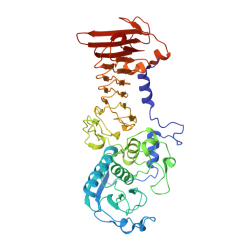
Crystal structure of a complex between Pseudomonas aeruginosa alkaline protease and its cognate inhibitor: inhibition by a zinc-NH2 coordinative bond

Hege, T.Feltzer, R.E.Gray, R.D.Baumann, U.

(2001) J Biol Chem 276: 35087-35092

  • DOI: https://doi.org/10.1074/jbc.M104020200
  • Primary Citation of Related Structures:  
    1GO7, 1GO8, 1JIW

  • PubMed Abstract: 

    Serralysins are a family of metalloproteases secreted by Gram-negative bacteria into the medium in the form of inactive zymogens. Usually, all serralysin secretors have on the same operon a gene coding for a periplasmic 10-kDa protein, which is an inhibitor of the secreted protease. The recent characterization of the inhibitor of the alkaline protease from Pseudomonas aeruginosa revealed a surprisingly low dissociation constant of 4 pm, contrary to earlier studies on homologous systems, where inhibition constants in the microm range were reported. To approach a more accurate understanding, the crystal structure of the complex between inhibitor and protease from P. aeruginosa was determined at 1.74 A resolution and refined to R(free) = 0.204. The structure reported here shows clearly that the N terminus of the inhibitor forms a coordinative bond to the catalytic Zn(2+) ion with a nitrogen-zinc distance of 2.17 A. We conclude that this interaction adds substantially to the complex stability and show also that similar interactions are found in other metzincin-inhibitor complexes.


  • Organizational Affiliation

    Department of Chemistry and Biochemistry, University of Bern, 3012 Bern, Switzerland.


Macromolecules
Find similar proteins by:  (by identity cutoff)  |  3D Structure
Entity ID: 1
MoleculeChains Sequence LengthOrganismDetailsImage
ALKALINE METALLOPROTEINASEA [auth P]470Pseudomonas aeruginosaMutation(s): 0 
EC: 3.4.24
UniProt
Find proteins for Q03023 (Pseudomonas aeruginosa (strain ATCC 15692 / DSM 22644 / CIP 104116 / JCM 14847 / LMG 12228 / 1C / PRS 101 / PAO1))
Explore Q03023 
Go to UniProtKB:  Q03023
Entity Groups  
Sequence Clusters30% Identity50% Identity70% Identity90% Identity95% Identity100% Identity
UniProt GroupQ03023
Sequence Annotations
Expand
  • Reference Sequence
Find similar proteins by:  (by identity cutoff)  |  3D Structure
Entity ID: 2
MoleculeChains Sequence LengthOrganismDetailsImage
PROTEINASE INHIBITORB [auth I]106Pseudomonas aeruginosaMutation(s): 0 
UniProt
Find proteins for Q03026 (Pseudomonas aeruginosa (strain ATCC 15692 / DSM 22644 / CIP 104116 / JCM 14847 / LMG 12228 / 1C / PRS 101 / PAO1))
Explore Q03026 
Go to UniProtKB:  Q03026
Entity Groups  
Sequence Clusters30% Identity50% Identity70% Identity90% Identity95% Identity100% Identity
UniProt GroupQ03026
Sequence Annotations
Expand
  • Reference Sequence
Experimental Data & Validation

Experimental Data

  • Method: X-RAY DIFFRACTION
  • Resolution: 1.74 Å
  • R-Value Free: 0.204 
  • R-Value Work: 0.184 
  • R-Value Observed: 0.185 
  • Space Group: P 21 21 2
Unit Cell:
Length ( Å )Angle ( ˚ )
a = 75.627α = 90
b = 118.432β = 90
c = 91.995γ = 90
Software Package:
Software NamePurpose
CNSrefinement
DENZOdata reduction
SCALEPACKdata scaling
CNSphasing

Structure Validation

View Full Validation Report


Entry History 

Deposition Data

Revision History  (Full details and data files)

  • Version 1.0: 2001-08-15
    Type: Initial release
  • Version 1.1: 2008-04-27
    Changes: Version format compliance
  • Version 1.2: 2011-07-13
    Changes: Version format compliance
  • Version 1.3: 2023-08-16
    Changes: Data collection, Database references, Derived calculations, Refinement description