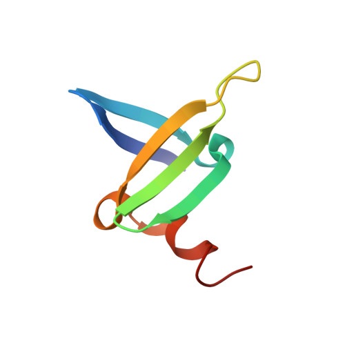
A single-point mutation in the extreme heat- and pressure-resistant sso7d protein from sulfolobus solfataricus leads to a major rearrangement of the hydrophobic core.
Consonni, R., Santomo, L., Fusi, P., Zetta, L.(1999) Biochemistry 38: 12709-12717
- PubMed: 10504241
- DOI: https://doi.org/10.1021/bi9911280
- Primary Citation of Related Structures:
1B4O, 1JIC - PubMed Abstract:
Sso7d is a basic 7-kDa DNA-binding protein from Sulfolobus solfataricus, also endowed with ribonuclease activity. The protein consists of a double-stranded antiparallel beta-sheet, onto which an orthogonal triple-stranded antiparallel beta-sheet is packed, and of a small helical stretch at the C-terminus. Furthermore, the two beta-sheets enclose an aromatic cluster displaying a fishbone geometry. We previously cloned the Sso7d-encoding gene, expressed it in Escherichia coli, and produced several single-point mutants, either of residues located in the hydrophobic core or of Trp23, which is exposed to the solvent and plays a major role in DNA binding. The mutation F31A was dramatically destabilizing, with a loss in thermo- and piezostabilities by at least 27 K and 10 kbar, respectively. Here, we report the solution structure of the F31A mutant, which was determined by NMR spectroscopy using 744 distance constraints obtained from analysis of multidimensional spectra in conjunction with simulated annealing protocols. The most remarkable finding is the change in orientation of the Trp23 side chain, which in the wild type is completely exposed to the solvent, whereas in the mutant is largely buried in the aromatic cluster. This prevents the formation of a cavity in the hydrophobic core of the mutant, which would arise in the absence of structural rearrangements. We found additional changes produced by the mutation, notably a strong distortion in the beta-sheets with loss in several hydrogen bonds, increased flexibility of some stretches of the backbone, and some local strains. On one hand, these features may justify the dramatic destabilization provoked by the mutation; on the other hand, they highlight the crucial role of the hydrophobic core in protein stability. To the best of our knowledge, no similar rearrangement has been so far described as a result of a single-point mutation.
Organizational Affiliation:
Istituto di Chimica delle Macromolecole, Lab. NMR, CNR, Via Ampère 56, 20131 Milano, Italy, and Dipartimento di BioTecnologie e Bioscienze, Università di Milano-Bicocca, Pza delle Scienze 2, 20126 Milano, Italy. roberto@labnmr.icmnmr.mi.cnr.it


