NMR derived solution structure of an EF-hand calcium-binding protein from Entamoeba Histolytica.
Atreya, H.S., Sahu, S.C., Bhattacharya, A., Chary, K.V., Govil, G.(2001) Biochemistry 40: 14392-14403
- PubMed: 11724551
- DOI: https://doi.org/10.1021/bi0114978
- Primary Citation of Related Structures:
1JFJ, 1JFK - PubMed Abstract:
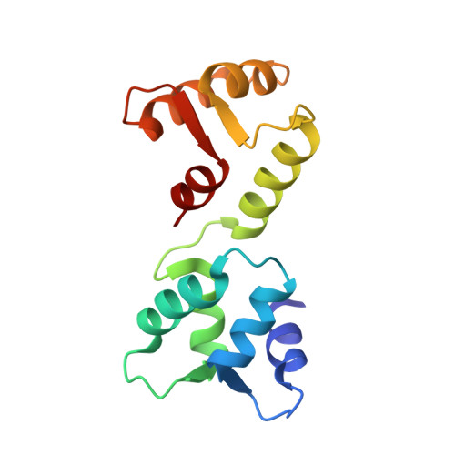
We present the three-dimensional (3D) solution structure of a calcium-binding protein from Entamoeba histolytica (EhCaBP), an etiologic agent of amoebiasis affecting millions worldwide. EhCaBP is a 14.7 kDa (134 residues) monomeric protein thought to play a role in the pathogenesis of amoebiasis. The 3D structure of Ca(2+)-bound EhCaBP has been derived using multidimensional nuclear magnetic resonance (NMR) spectroscopic techniques. The study reveals the presence of two globular domains connected by a flexible linker region spanning 8 amino acid residues. Each domain consists of a pair of helix-loop-helix motifs similar to the canonical EF-hand motif of calcium-binding proteins. EhCaBP binds to four Ca(2+) with high affinity (two in each domain), and it is structurally related to calmodulin (CaM) and troponin C (TnC) despite its low sequence homology ( approximately 29%) with these proteins. NMR-derived structures of EhCaBP converge within each domain with low RMSDs and angular order-parameters for backbone torsion angles close to 1.0. However, the presence of a highly flexible central linker region results in an ill-defined orientation of the two domains relative to one other. These findings are supported by backbone (15)N relaxation rate measurements and deuterium exchange studies, which reveal low structural order parameters for residues in the central linker region. Earlier, biochemical studies showed that EhCaBP is involved in a novel signal transduction mechanism, distinct from CaM. A possible reason for such a functional diversity is revealed by a detailed comparison of the 3D structure of EhCaBP with that of CaM and TnC. The studies indicate a more open C-terminal domain for EhCaBP with larger water exposed total hydrophobic surface area as compared to CaM and TnC. Further dissimilarities between the structures include the presence of two Gly residues (G63 and G67) in the central linker region of EhCaBP, which seem to impart it a greater flexibility compared to CaM and TnC and also play crucial role in its biological function. Thus, unlike in CaM and TnC, wherein the length and/or composition of the central linker have been found to be crucial for their function, in EhCaBP, both flexibility as well as amino acid composition is required for the function of the protein.
Organizational Affiliation:
Department of Chemical Sciences, Tata Institute of Fundamental Research, Mumbai-400005, India.