1JFH

STRUCTURE OF A PANCREATIC ALPHA-AMYLASE BOUND TO A SUBSTRATE ANALOGUE AT 2.03 ANGSTROM RESOLUTION


Experimental Data Snapshot

  • Method: X-RAY DIFFRACTION
  • Resolution: 2.03 Å
  • R-Value Free: 0.185 
  • R-Value Work: 0.160 
  • R-Value Observed: 0.160 

wwPDB Validation   3D Report Full Report


This is version 3.0 of the entry. See complete history


Literature

Structure of a pancreatic alpha-amylase bound to a substrate analogue at 2.03 A resolution.

Qian, M.Spinelli, S.Driguez, H.Payan, F.

(1997) Protein Sci 6: 2285-2296

  • DOI: https://doi.org/10.1002/pro.5560061102
  • Primary Citation of Related Structures:  
    1JFH

  • PubMed Abstract: 

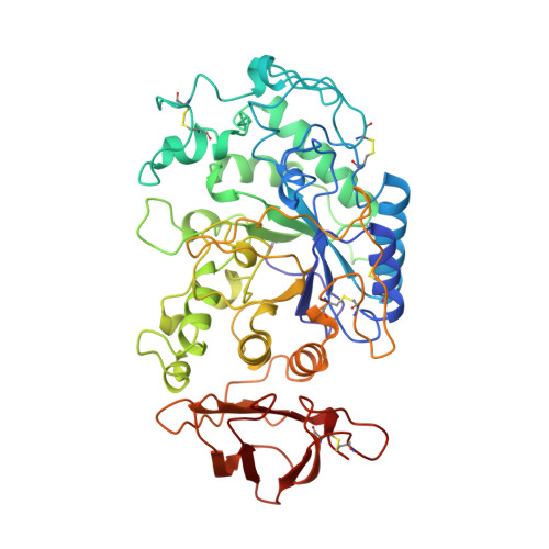
    The structure of pig pancreatic alpha-amylase in complex with carbohydrate inhibitor and proteinaceous inhibitors is known but the successive events occurring at the catalytic center still remain to be elucidated. The X-ray structure analysis of a crystal of pig pancreatic alpha-amylase (PPA, EC 3.2.1.1.) soaked with an enzyme-resistant substrate analogue, methyl 4,4'-dithio-alpha-maltotrioside, showed electron density corresponding to the binding of substrate analogue molecules at the active site and at the "second binding site." The electron density observed at the active site was interpreted in terms of overlapping networks of oligosaccharides, which show binding of substrate analogue molecules at subsites prior to and subsequent to the cleavage site. A weaker patch of density observed at subsite -1 (using a nomenclature where the site of hydrolysis is taken to be between subsites -1 and +1) was modeled with water molecules. Conformational changes take place upon substrate analogue binding and the "flexible loop" that constitutes the surface edge of the active site is observed in a specific conformation. This confirms that this loop plays an important role in the recognition and binding of the ligand. The crystal structure was refined at 2.03 A resolution, to an R-factor of 16.0 (Rfree, 18.5).


  • Organizational Affiliation

    AFMB-IBSM-CNRS, Marseille, France.


Macromolecules
Find similar proteins by:  (by identity cutoff)  |  3D Structure
Entity ID: 1
MoleculeChains Sequence LengthOrganismDetailsImage
ALPHA-AMYLASE496Sus scrofaMutation(s): 0 
EC: 3.2.1.1
UniProt
Find proteins for P00690 (Sus scrofa)
Explore P00690 
Go to UniProtKB:  P00690
Entity Groups  
Sequence Clusters30% Identity50% Identity70% Identity90% Identity95% Identity100% Identity
UniProt GroupP00690
Sequence Annotations
Expand
  • Reference Sequence
Oligosaccharides

Help

Entity ID: 2
MoleculeChains Length2D Diagram Glycosylation3D Interactions
4-S-methyl-4-thio-alpha-D-glucopyranose-(1-4)-methyl 4-thio-alpha-D-glucopyranoside
B
2N/AN/A
Glycosylation Resources
GlyTouCan:  G59184PB
GlyCosmos:  G59184PB
Entity ID: 3
MoleculeChains Length2D Diagram Glycosylation3D Interactions
alpha-D-glucopyranose-(1-4)-1,4-dithio-alpha-D-glucopyranose
C, D
2N/AN/A
Glycosylation Resources
GlyTouCan:  G11151AA
GlyCosmos:  G11151AA
Small Molecules
Ligands 3 Unique
IDChains Name / Formula / InChI Key2D Diagram3D Interactions
HG
Query on HG

Download Ideal Coordinates CCD File 
F [auth A]MERCURY (II) ION
Hg
BQPIGGFYSBELGY-UHFFFAOYSA-N
CA
Query on CA

Download Ideal Coordinates CCD File 
G [auth A]CALCIUM ION
Ca
BHPQYMZQTOCNFJ-UHFFFAOYSA-N
CL
Query on CL

Download Ideal Coordinates CCD File 
E [auth A]CHLORIDE ION
Cl
VEXZGXHMUGYJMC-UHFFFAOYSA-M
Modified Residues  1 Unique
IDChains TypeFormula2D DiagramParent
PCA
Query on PCA
A
L-PEPTIDE LINKINGC5 H7 N O3GLN
Experimental Data & Validation

Experimental Data

  • Method: X-RAY DIFFRACTION
  • Resolution: 2.03 Å
  • R-Value Free: 0.185 
  • R-Value Work: 0.160 
  • R-Value Observed: 0.160 
  • Space Group: P 21 21 21
Unit Cell:
Length ( Å )Angle ( ˚ )
a = 56.3α = 90
b = 87.8β = 90
c = 103.4γ = 90
Software Package:
Software NamePurpose
DENZOdata reduction
X-PLORmodel building
X-PLORrefinement
X-PLORphasing

Structure Validation

View Full Validation Report


Entry History 

Deposition Data

Revision History  (Full details and data files)

  • Version 1.0: 1998-12-02
    Type: Initial release
  • Version 1.1: 2008-03-24
    Changes: Version format compliance
  • Version 1.2: 2011-07-13
    Changes: Version format compliance
  • Version 2.0: 2019-12-25
    Changes: Data collection, Database references, Derived calculations, Polymer sequence
  • Version 3.0: 2020-07-29
    Type: Remediation
    Reason: Carbohydrate remediation
    Changes: Atomic model, Data collection, Derived calculations, Structure summary