1JEB

Chimeric Human/Mouse Carbonmonoxy Hemoglobin (Human Zeta2 / Mouse Beta2)


Experimental Data Snapshot

  • Method: X-RAY DIFFRACTION
  • Resolution: 2.10 Å
  • R-Value Free: 0.249 
  • R-Value Work: 0.216 

wwPDB Validation   3D Report Full Report


Ligand Structure Quality Assessment 


This is version 1.3 of the entry. See complete history


Literature

The role of beta chains in the control of the hemoglobin oxygen binding function: chimeric human/mouse proteins, structure, and function.

Kidd, R.D.Russell, J.E.Watmough, N.J.Baker, E.N.Brittain, T.

(2001) Biochemistry 40: 15669-15675

  • DOI: https://doi.org/10.1021/bi011329f
  • Primary Citation of Related Structures:  
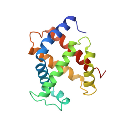
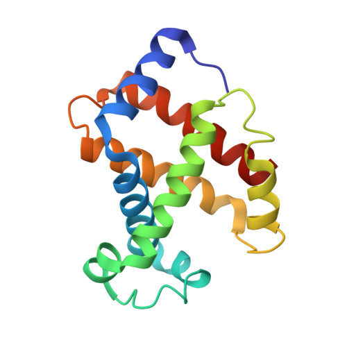
    1JEB

  • PubMed Abstract: 

    By using transgenic methodologies, we have produced a number of mouse/human chimeric hemoglobins containing adult mouse and human embryonic globin chains. A detailed analysis of the oxygen binding properties of these proteins identifies the dominant role played by the specific beta-type globin chains in the control of the oxygen binding characteristics. Further analysis traces the origins of these effects to alterations in the properties of the T states of these proteins. The human zeta/mouse beta chimeric protein has been crystallized, and its structure has been determined by X-ray diffraction to a resolution of 2.1 A with R (R(free)) values of 21.6% (24.9%). Close examination of the structure indicates that the subunit interfaces contain contacts which, although different from those present in either the parent human or the parent mouse proteins, retain the overall stabilizing interactions seen in other R state hemoglobins.


  • Organizational Affiliation

    School of Biological Sciences, Private Bag 92019, University of Auckland, Auckland, New Zealand.


Macromolecules
Find similar proteins by:  (by identity cutoff)  |  3D Structure
Entity ID: 1
MoleculeChains Sequence LengthOrganismDetailsImage
HEMOGLOBIN ZETA CHAIN
A, C
142Homo sapiensMutation(s): 0 
Gene Names: HBZ
UniProt & NIH Common Fund Data Resources
Find proteins for P02008 (Homo sapiens)
Explore P02008 
Go to UniProtKB:  P02008
PHAROS:  P02008
GTEx:  ENSG00000130656 
Entity Groups  
Sequence Clusters30% Identity50% Identity70% Identity90% Identity95% Identity100% Identity
UniProt GroupP02008
Sequence Annotations
Expand
  • Reference Sequence
Find similar proteins by:  (by identity cutoff)  |  3D Structure
Entity ID: 2
MoleculeChains Sequence LengthOrganismDetailsImage
HEMOGLOBIN BETA-SINGLE CHAIN
B, D
146Mus musculusMutation(s): 0 
UniProt
Find proteins for P02088 (Mus musculus)
Explore P02088 
Go to UniProtKB:  P02088
Entity Groups  
Sequence Clusters30% Identity50% Identity70% Identity90% Identity95% Identity100% Identity
UniProt GroupP02088
Sequence Annotations
Expand
  • Reference Sequence
Experimental Data & Validation

Experimental Data

  • Method: X-RAY DIFFRACTION
  • Resolution: 2.10 Å
  • R-Value Free: 0.249 
  • R-Value Work: 0.216 
  • Space Group: P 41
Unit Cell:
Length ( Å )Angle ( ˚ )
a = 84.921α = 90
b = 84.921β = 90
c = 104.523γ = 90
Software Package:
Software NamePurpose
CNSrefinement
ADSCdata collection
DENZOdata reduction
SCALEPACKdata scaling
CNSphasing

Structure Validation

View Full Validation Report


Ligand Structure Quality Assessment 


Entry History 

Deposition Data

Revision History  (Full details and data files)

  • Version 1.0: 2002-01-23
    Type: Initial release
  • Version 1.1: 2008-04-27
    Changes: Version format compliance
  • Version 1.2: 2011-07-13
    Changes: Version format compliance
  • Version 1.3: 2023-08-16
    Changes: Data collection, Database references, Derived calculations, Refinement description