1JE5

Crystal Structure of gp2.5, a Single-Stranded DNA Binding Protein Encoded by Bacteriophage T7


Experimental Data Snapshot

  • Method: X-RAY DIFFRACTION
  • Resolution: 1.90 Å
  • R-Value Free: 0.268 
  • R-Value Work: 0.221 

wwPDB Validation   3D Report Full Report


This is version 1.3 of the entry. See complete history


Literature

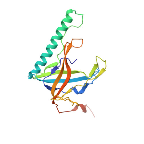
Structure of the gene 2.5 protein, a single-stranded DNA binding protein encoded by bacteriophage T7.

Hollis, T.Stattel, J.M.Walther, D.S.Richardson, C.C.Ellenberger, T.

(2001) Proc Natl Acad Sci U S A 98: 9557-9562

  • DOI: https://doi.org/10.1073/pnas.171317698
  • Primary Citation of Related Structures:  
    1JE5

  • PubMed Abstract: 

    The gene 2.5 protein (gp2.5) of bacteriophage T7 is a single-stranded DNA (ssDNA) binding protein that has essential roles in DNA replication and recombination. In addition to binding DNA, gp2.5 physically interacts with T7 DNA polymerase and T7 primase-helicase during replication to coordinate events at the replication fork. We have determined a 1.9-A crystal structure of gp2.5 and show that it has a conserved OB-fold (oligosaccharide/oligonucleotide binding fold) that is well adapted for interactions with ssDNA. Superposition of the OB-folds of gp2.5 and other ssDNA binding proteins reveals a conserved patch of aromatic residues that stack against the bases of ssDNA in the other crystal structures, suggesting that gp2.5 binds to ssDNA in a similar manner. An acidic C-terminal extension of the gp2.5 protein, which is required for dimer formation and for interactions with the T7 DNA polymerase and the primase-helicase, appears to be flexible and may act as a switch that modulates the DNA binding affinity of gp2.5.


  • Organizational Affiliation

    Department of Biological Chemistry and Molecular Pharmacology, Harvard Medical School, Boston, MA 02115, USA.


Macromolecules
Find similar proteins by:  (by identity cutoff)  |  3D Structure
Entity ID: 1
MoleculeChains Sequence LengthOrganismDetailsImage
HELIX-DESTABILIZING PROTEIN
A, B
206Escherichia phage T7Mutation(s): 0 
UniProt
Find proteins for P03696 (Escherichia phage T7)
Explore P03696 
Go to UniProtKB:  P03696
Entity Groups  
Sequence Clusters30% Identity50% Identity70% Identity90% Identity95% Identity100% Identity
UniProt GroupP03696
Sequence Annotations
Expand
  • Reference Sequence
Small Molecules
Ligands 1 Unique
IDChains Name / Formula / InChI Key2D Diagram3D Interactions
CA
Query on CA

Download Ideal Coordinates CCD File 
C [auth B]CALCIUM ION
Ca
BHPQYMZQTOCNFJ-UHFFFAOYSA-N
Experimental Data & Validation

Experimental Data

  • Method: X-RAY DIFFRACTION
  • Resolution: 1.90 Å
  • R-Value Free: 0.268 
  • R-Value Work: 0.221 
  • Space Group: P 21 21 21
Unit Cell:
Length ( Å )Angle ( ˚ )
a = 68.1α = 90
b = 71.8β = 90
c = 82.2γ = 90
Software Package:
Software NamePurpose
MLPHAREphasing
CNSrefinement
DENZOdata reduction
SCALEPACKdata scaling

Structure Validation

View Full Validation Report


Entry History 

Deposition Data

Revision History  (Full details and data files)

  • Version 1.0: 2001-08-22
    Type: Initial release
  • Version 1.1: 2008-04-27
    Changes: Version format compliance
  • Version 1.2: 2011-07-13
    Changes: Version format compliance
  • Version 1.3: 2024-02-07
    Changes: Data collection, Database references, Derived calculations