1J80

Osmolyte Stabilization of RNase


Experimental Data Snapshot

  • Method: X-RAY DIFFRACTION
  • Resolution: 2.10 Å
  • R-Value Free: 0.282 
  • R-Value Work: 0.218 
  • R-Value Observed: 0.218 

wwPDB Validation   3D Report Full Report


This is version 1.3 of the entry. See complete history


Literature

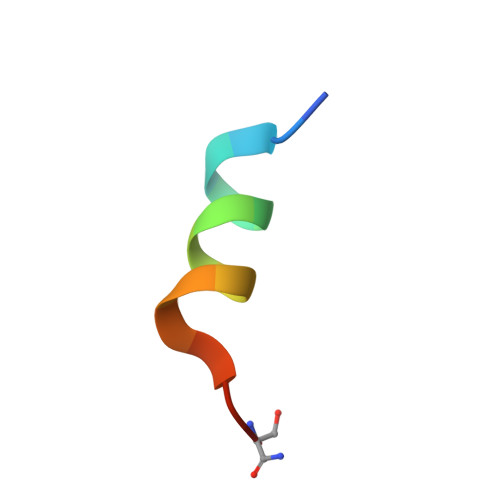
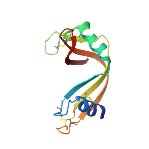
Osmolytes stabilize ribonuclease S by stabilizing its fragments S protein and S peptide to compact folding-competent states.

Ratnaparkhi, G.S.Varadarajan, R.

(2001) J Biol Chem 276: 28789-28798

  • DOI: https://doi.org/10.1074/jbc.M101906200
  • Primary Citation of Related Structures:  
    1J7Z, 1J80, 1J81, 1J82

  • PubMed Abstract: 

    Osmolytes stabilize proteins to thermal and chemical denaturation. We have studied the effects of the osmolytes sarcosine, betaine, trimethylamine-N-oxide, and taurine on the structure and stability of the protein.peptide complex RNase S using x-ray crystallography and titration calorimetry, respectively. The largest degree of stabilization is achieved with 6 m sarcosine, which increases the denaturation temperatures of RNase S and S pro by 24.6 and 17.4 degrees C, respectively, at pH 5 and protects both proteins against tryptic cleavage. Four crystal structures of RNase S in the presence of different osmolytes do not offer any evidence for osmolyte binding to the folded state of the protein or any perturbation in the water structure surrounding the protein. The degree of stabilization in 6 m sarcosine increases with temperature, ranging from -0.52 kcal mol(-1) at 20 degrees C to -5.4 kcal mol(-1) at 60 degrees C. The data support the thesis that osmolytes that stabilize proteins, do so by perturbing unfolded states, which change conformation to a compact, folding competent state in the presence of osmolyte. The increased stabilization thus results from a decrease in conformational entropy of the unfolded state.


  • Organizational Affiliation

    National Center for Biological Sciences, Bangalore 560 065, India.


Macromolecules

Find similar proteins by:  Sequence   |   3D Structure  

Entity ID: 1
MoleculeChains Sequence LengthOrganismDetailsImage
RIBONUCLEASE PANCREATIC16N/AMutation(s): 1 
EC: 3.1.27.5
UniProt
Find proteins for P61823 (Bos taurus)
Explore P61823 
Go to UniProtKB:  P61823
Entity Groups  
Sequence Clusters30% Identity50% Identity70% Identity90% Identity95% Identity100% Identity
UniProt GroupP61823
Sequence Annotations
Expand
  • Reference Sequence
Find similar proteins by:  (by identity cutoff)  |  3D Structure
Entity ID: 2
MoleculeChains Sequence LengthOrganismDetailsImage
RIBONUCLEASE PANCREATIC104Bos taurusMutation(s): 0 
EC: 3.1.27.5
UniProt
Find proteins for P61823 (Bos taurus)
Explore P61823 
Go to UniProtKB:  P61823
Entity Groups  
Sequence Clusters30% Identity50% Identity70% Identity90% Identity95% Identity100% Identity
UniProt GroupP61823
Sequence Annotations
Expand
  • Reference Sequence
Small Molecules
Ligands 1 Unique
IDChains Name / Formula / InChI Key2D Diagram3D Interactions
SO4
Query on SO4

Download Ideal Coordinates CCD File 
C [auth B]SULFATE ION
O4 S
QAOWNCQODCNURD-UHFFFAOYSA-L
Experimental Data & Validation

Experimental Data

  • Method: X-RAY DIFFRACTION
  • Resolution: 2.10 Å
  • R-Value Free: 0.282 
  • R-Value Work: 0.218 
  • R-Value Observed: 0.218 
  • Space Group: P 31 2 1
Unit Cell:
Length ( Å )Angle ( ˚ )
a = 44.6α = 90
b = 44.6β = 90
c = 98.14γ = 120
Software Package:
Software NamePurpose
XDSdata scaling
XDSdata reduction
X-PLORmodel building
X-PLORrefinement
X-PLORphasing

Structure Validation

View Full Validation Report


Entry History 

Deposition Data

Revision History  (Full details and data files)

  • Version 1.0: 2001-06-06
    Type: Initial release
  • Version 1.1: 2008-04-27
    Changes: Version format compliance
  • Version 1.2: 2011-07-13
    Changes: Version format compliance
  • Version 1.3: 2017-10-04
    Changes: Refinement description