Crystal Structure of Thermostable Aspartase from Bacillus sp. YM55-1: Structure-based Exploration of Functional Sites in the Aspartase Family
Fujii, T., Sakai, H., Kawata, Y., Hata, Y.(2003) J Mol Biol 328: 635-654
- PubMed: 12706722
- DOI: https://doi.org/10.1016/s0022-2836(03)00310-3
- Primary Citation of Related Structures:
1J3U - PubMed Abstract:
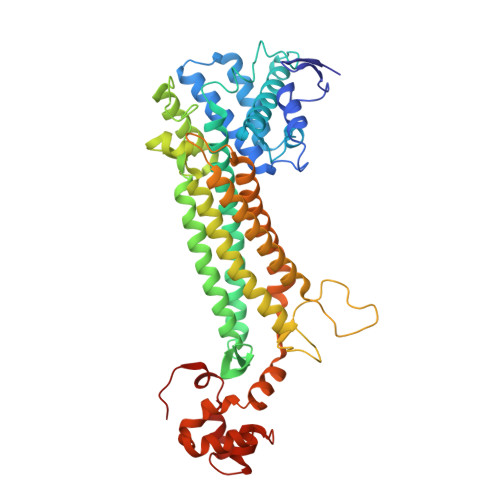
The crystal structure of the thermostable aspartase from Bacillus sp. YM55-1 has been solved and refined for 2.5A resolution data with an R-factor of 22.1%. The present enzyme is a homotetramer with subunits composed of three domains. It exhibits no allosteric effects, in contrast to the Escherichia coli aspartase, which is activated by divalent metal cation and L-aspartate, but is four-times more active than the E.coli enzyme. The overall folding of the present enzyme subunit is similar to those of the E.coli aspartase and the E.coli fumarase C, both of which belong to the same superfamily as the present enzyme. A local structural comparison of these three enzymes revealed seven structurally different regions. Five of the regions were located around putative functional sites, suggesting the involvement of these regions into the functions characteristic of the enzymes. Of these regions, the region of Gln96-Gly100 is proposed as a part of the recognition site of the alpha-amino group in L-aspartate for aspartase and the hydroxyl group in L-malate for fumarase. The region of Gln315-Gly323 is a flexible loop with a well-conserved sequence that is suggested to be involved in the catalytic reaction. The region of Lys123-Lys128 corresponds to a part of the putative activator-binding site in the E.coli fumarase C. The region in the Bacillus aspartase, however, adopts a main-chain conformation that prevents the activator binding. The regions of Gly228-Glu241 and Val265-Asp272, which form a part of the active-site wall, are suggested to be involved in the allosteric activation of the E.coli aspartase by the binding of the metal ion and the activator. Moreover, an increase in the numbers of intersubunit hydrogen bonds and salt-bridges is observed in the Bacillus aspartase relative to those of the E.coli enzyme, implying a contribution to the thermostability of the present aspartase.
Organizational Affiliation:
Institute for Chemical Research, Kyoto University, Uji, Kyoto 611-0011, Japan.