


1J2E
Crystal structure of Human Dipeptidyl peptidase IV
- PDB DOI: https://doi.org/10.2210/pdb1J2E/pdb
- Classification: HYDROLASE
- Organism(s): Homo sapiens
- Expression System: Spodoptera frugiperda
- Mutation(s): No
- Deposited: 2002-12-30 Released: 2003-12-30
Experimental Data Snapshot
- Method: X-RAY DIFFRACTION
- Resolution: 2.60 Å
- R-Value Free: 0.302
- R-Value Work: 0.249
wwPDB Validation 3D Report Full Report

This is version 1.4 of the entry. See complete history.

Macromolecules
Find similar proteins by:
(by identity cutoff) | 3D Structure
Entity ID: 1 | |||||
|---|---|---|---|---|---|
| Molecule | Chains | Sequence Length | Organism | Details | Image |
| Dipeptidyl peptidase IV | 740 | Homo sapiens | Mutation(s): 0 EC: 3.4.14.5 | 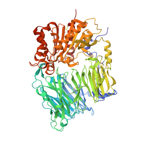 | |
UniProt & NIH Common Fund Data Resources | |||||
Find proteins for P27487 (Homo sapiens) Explore P27487 Go to UniProtKB: P27487 | |||||
PHAROS: P27487 GTEx: ENSG00000197635 | |||||
Entity Groups | |||||
| Sequence Clusters | 30% Identity50% Identity70% Identity90% Identity95% Identity100% Identity | ||||
| UniProt Group | P27487 | ||||
Sequence AnnotationsExpand | |||||
| |||||
Small Molecules
| Ligands 1 Unique | |||||
|---|---|---|---|---|---|
| ID | Chains | Name / Formula / InChI Key | 2D Diagram | 3D Interactions | |
| NAG Query on NAG | C [auth A] D [auth A] E [auth A] F [auth A] G [auth A] | 2-acetamido-2-deoxy-beta-D-glucopyranose C8 H15 N O6 OVRNDRQMDRJTHS-FMDGEEDCSA-N |  | ||
Experimental Data & Validation
Experimental Data
- Method: X-RAY DIFFRACTION
- Resolution: 2.60 Å
- R-Value Free: 0.302
- R-Value Work: 0.249
- Space Group: P 21 21 21
Unit Cell:
| Length ( Å ) | Angle ( ˚ ) |
|---|---|
| a = 118.04 | α = 90 |
| b = 125.92 | β = 90 |
| c = 136.84 | γ = 90 |
| Software Name | Purpose |
|---|---|
| MOSFLM | data reduction |
| SCALA | data scaling |
| MLPHARE | phasing |
| CNX | refinement |
| CCP4 | data scaling |
Entry History
Deposition Data
- Released Date: 2003-12-30 Deposition Author(s): Hiramatsu, H., Kyono, K., Higashiyama, Y., Fukushima, C., Shima, H., Sugiyama, S., Inaka, K., Yamamoto, A., Shimizu, R.
Revision History (Full details and data files)
- Version 1.0: 2003-12-30
Type: Initial release - Version 1.1: 2008-04-27
Changes: Version format compliance - Version 1.2: 2011-07-13
Changes: Non-polymer description, Version format compliance - Version 1.3: 2020-07-29
Type: Remediation
Reason: Carbohydrate remediation
Changes: Data collection, Database references, Derived calculations, Structure summary - Version 1.4: 2023-12-27
Changes: Data collection, Database references, Structure summary
