Crystal Structure of Extended-Spectrum beta-Lactamase Toho-1: Insights into the Molecular Mechanism for Catalytic Reaction and Substrate Specificity Expansion
Ibuka, A.S., Ishii, Y., Galleni, M., Ishiguro, M., Yamaguchi, K., Frere, J.M., Matsuzawa, H., Sakai, H.(2003) Biochemistry 42: 10634-10643
- PubMed: 12962487
- DOI: https://doi.org/10.1021/bi0342822
- Primary Citation of Related Structures:
1IYS - PubMed Abstract:
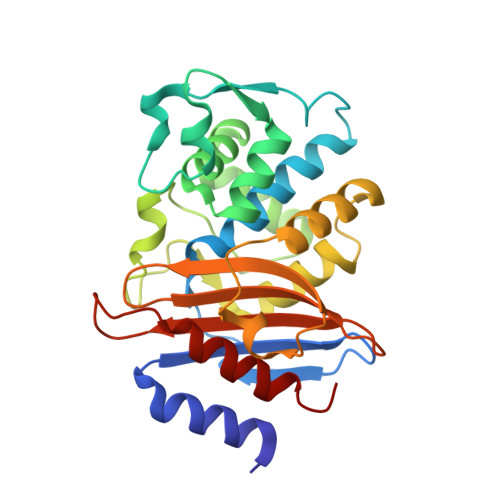
The crystallographic structure of the class A beta-lactamase Toho-1, an extended-spectrum beta-lactamase with potent activity against expanded-spectrum cephems, has been determined at 1.65 A resolution. The result reveals that the Lys73 side chain can adopt two alternative conformations. The predominant conformation of Lys73 is different from that observed in the E166A mutant, indicating that removal of the Glu166 side chain changes the conformation of the Lys73 side chain and thus the interaction between Lys73 and Glu166. The Lys73 side chain would play an important role in proton relay, switching its conformation from one to the other depending on the circumstances. The electron density map also implies possible rotation of Ser237. Comparison of the Toho-1 structure with the structure of other class A beta-lactamases shows that the hydroxyl group of Ser237 is likely to rotate through interaction with the carboxyl group of the substrate. Another peculiarity is the existence of three sulfate ions positioned in or near the substrate-binding cavity. One of these sulfate ions is tightly bound to the active center, while the other two are held by a region of positive charge formed by two arginine residues, Arg274 and Arg276. This positively charged region is speculated to represent a pseudo-binding site of the beta-lactam antibiotics, presumably catching the methoxyimino group of the third-generation cephems prior to proper binding in the substrate-binding cleft for hydrolysis. This high-resolution structure, together with detailed kinetic analysis of Toho-1, provides a new hypothesis for the catalytic mechanism and substrate specificity of Toho-1.
Organizational Affiliation:
Department of Food and Nutritional Sciences, University of Shizuoka, 52-1 Yada, Shizuoka 422-8526, Japan. ibuka@u-shizuoka-ken.ac.jp