1IIQ

CRYSTAL STRUCTURE OF HIV-1 PROTEASE COMPLEXED WITH A HYDROXYETHYLAMINE PEPTIDOMIMETIC INHIBITOR


Experimental Data Snapshot

  • Method: X-RAY DIFFRACTION
  • Resolution: 1.83 Å
  • R-Value Free: 0.204 
  • R-Value Work: 0.164 
  • R-Value Observed: 0.164 

wwPDB Validation   3D Report Full Report


This is version 1.6 of the entry. See complete history


Literature

Hydroxyethylamine isostere of an HIV-1 protease inhibitor prefers its amine to the hydroxy group in binding to catalytic aspartates. A synchrotron study of HIV-1 protease in complex with a peptidomimetic inhibitor.

Dohnalek, J.Hasek, J.Duskova, J.Petrokova, H.Hradilek, M.Soucek, M.Konvalinka, J.Brynda, J.Sedlacek, J.Fabry, M.

(2002) J Med Chem 45: 1432-1438

  • DOI: https://doi.org/10.1021/jm010979e
  • Primary Citation of Related Structures:  
    1IIQ

  • PubMed Abstract: 

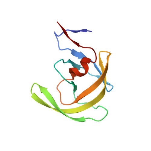
    A complex structure of HIV-1 protease with a hydroxyethylamine-containing inhibitor Boc-Phe-Psi[(S)-CH(OH)CH2NH]-Phe-Gln-Phe-NH2 has been determined by X-ray diffraction to 1.8 A resolution. The inhibitor is bound in the active site of the protease dimer with its hydroxyethylamine isostere participating in hydrogen bonds to the catalytic aspartates 25 and 25' and glycine 27' of the active site triads via five hydrogen bonds. The isostere amine interactions with the catalytic aspartates result in a displacement of the isostere hydroxy group in comparison with the common position known for analogous hydroxyethylamine containing inhibitors. A comparison with another inhibitor of this series shows that the change of one atom of the P2' side chain (Glu/Gln) leads to an altered ability of creating hydrogen bonds to the active site and within the inhibitor molecule. The diffraction data collected at a synchrotron radiation source enabled a detailed analysis of the complex solvation and of alternative conformations of protein side chains.


  • Organizational Affiliation

    Institute of Molecular Genetics, Academy of Sciences of the Czech Republic, Flemingovo nám. 2, 166 10 Praha 6, Czech Republic. dohnalek@imc.cas.cz


Macromolecules
Find similar proteins by:  (by identity cutoff)  |  3D Structure
Entity ID: 1
MoleculeChains Sequence LengthOrganismDetailsImage
PROTEASE RETROPEPSIN
A, B
99Human immunodeficiency virus 1Mutation(s): 0 
EC: 3.4.23.16
UniProt
Find proteins for P03367 (Human immunodeficiency virus type 1 group M subtype B (isolate BRU/LAI))
Explore P03367 
Go to UniProtKB:  P03367
Entity Groups  
Sequence Clusters30% Identity50% Identity70% Identity90% Identity95% Identity100% Identity
UniProt GroupP03367
Sequence Annotations
Expand
  • Reference Sequence
Small Molecules
Binding Affinity Annotations 
IDSourceBinding Affinity
0ZR BindingDB:  1IIQ Ki: min: 33, max: 1000 (nM) from 3 assay(s)
Binding MOAD:  1IIQ Ki: 33 (nM) from 1 assay(s)
Biologically Interesting Molecules (External Reference) 1 Unique
Experimental Data & Validation

Experimental Data

Unit Cell:
Length ( Å )Angle ( ˚ )
a = 58.11α = 90
b = 86.34β = 90
c = 46.11γ = 90
Software Package:
Software NamePurpose
AMoREphasing
CNSrefinement
XDSdata reduction
SCALEPACKdata scaling

Structure Validation

View Full Validation Report


Entry History 

Deposition Data

Revision History  (Full details and data files)

  • Version 1.0: 2002-04-12
    Type: Initial release
  • Version 1.1: 2007-10-16
    Changes: Version format compliance
  • Version 1.2: 2011-07-13
    Changes: Atomic model, Database references, Derived calculations, Non-polymer description, Structure summary, Version format compliance
  • Version 1.3: 2012-12-12
    Changes: Other
  • Version 1.4: 2017-10-04
    Changes: Advisory, Refinement description
  • Version 1.5: 2023-08-16
    Changes: Advisory, Data collection, Database references, Derived calculations, Refinement description
  • Version 1.6: 2024-03-13
    Changes: Source and taxonomy, Structure summary