1IHT

CRYSTAL STRUCTURE OF THE COMPLEX OF HUMAN ALPHA-THROMBIN AND NON-HYDROLYZABLE BIFUNCTIONAL INHIBITORS, HIRUTONIN-2 AND HIRUTONIN-6


Experimental Data Snapshot

  • Method: X-RAY DIFFRACTION
  • Resolution: 2.10 Å
  • R-Value Work: 0.162 
  • R-Value Observed: 0.162 

wwPDB Validation   3D Report Full Report


This is version 2.0 of the entry. See complete history


Literature

Crystal structure of the complex of human alpha-thrombin and nonhydrolyzable bifunctional inhibitors, hirutonin-2 and hirutonin-6.

Zdanov, A.Wu, S.DiMaio, J.Konishi, Y.Li, Y.Wu, X.Edwards, B.F.Martin, P.D.Cygler, M.

(1993) Proteins 17: 252-265

  • DOI: https://doi.org/10.1002/prot.340170304
  • Primary Citation of Related Structures:  
    1IHS, 1IHT

  • PubMed Abstract: 

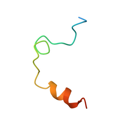
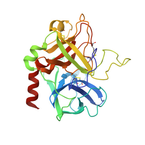
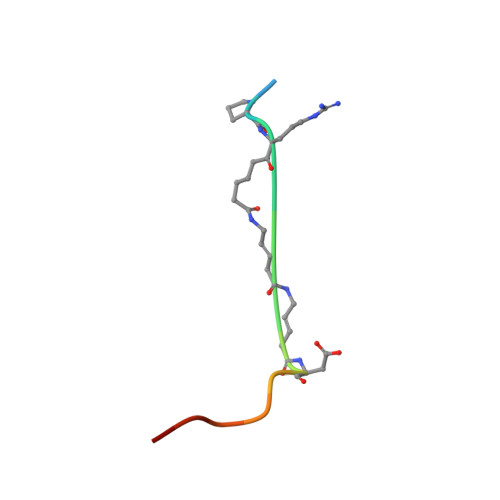
    The crystal structure of the complexes of hirutonin-2 and hirutonin-6 with human alpha-thrombin have been solved and refined to R-factors of 0.169 (2.0 A resolution) and 0.162 (2.1 A), respectively. Hirutonins belong to a family of bifunctional inhibitors bearing a noncleavable moiety mimicking the scissile bond. Hirutonin-2 is an analog of (D)Phe-Pro-Arg-Gly-hirudin49-65; hirutonin-6 has the same N-terminal tripeptide connected to a shortened fibrinogen exosite-binding part by a short, nonpeptidyl linker. The hirutonin-6 molecule is well defined in the electron density with the exception of the C-terminal Leu-h61. The linker follows near the bottom of the canyon connecting the active site with the exosite, forms a short antiparallel beta-sheet-like arrangement with Leu40-Leu41 and makes van der Waals contacts with Glu39-Leu40-Leu41 of thrombin. In the thrombin-hirutonin-2 complex, the N- and C-terminal parts of the inhibitor are well ordered (except the C-terminal Gln-h65) while the central portion of the linker is partially disordered. The glycine analog in the P1' position of hirutonin-2 assumes a conformation similar to that of the canonical form (Bode and Huber (1992) Eur. J. Biochem. 204:433-451) and supports the identification of the S1' site as restricted by His57, Trp60D, Lys60F, and the Cys42-Cys58 disulfide bridge. The carbonyl oxygen of the P1 arginine residue is located in the oxyanion hole formed by the NH groups of Gly193 and Ser195, while the carbonyl carbon is positioned within a short distance, 2.8 A, from the O gamma of Ser195. This resembles the conformation of the substrate-like inhibitors bound to other serine proteases. The N-terminal (D)Phe-Pro-Arg fragment common to both inhibitors binds to thrombin in a fashion very similar to that of other inhibitors having this motif. The binding of the C-terminus of hirutonins to the fibrinogen-binding exosite is similar to that observed in hirudin and hirulog complexes.


  • Organizational Affiliation

    Biotechnology Research Institute, National Research Council of Canada, Montréal, Québec.


Macromolecules
Find similar proteins by:  (by identity cutoff)  |  3D Structure
Entity ID: 1
MoleculeChains Sequence LengthOrganismDetailsImage
ALPHA-THROMBIN (SMALL SUBUNIT)A [auth L]36Homo sapiensMutation(s): 0 
EC: 3.4.21.5
UniProt & NIH Common Fund Data Resources
Find proteins for P00734 (Homo sapiens)
Explore P00734 
Go to UniProtKB:  P00734
PHAROS:  P00734
GTEx:  ENSG00000180210 
Entity Groups  
Sequence Clusters30% Identity50% Identity70% Identity90% Identity95% Identity100% Identity
UniProt GroupP00734
Sequence Annotations
Expand
  • Reference Sequence
Find similar proteins by:  (by identity cutoff)  |  3D Structure
Entity ID: 2
MoleculeChains Sequence LengthOrganismDetailsImage
ALPHA-THROMBIN (LARGE SUBUNIT)B [auth H]259Homo sapiensMutation(s): 0 
EC: 3.4.21.5
UniProt & NIH Common Fund Data Resources
Find proteins for P00734 (Homo sapiens)
Explore P00734 
Go to UniProtKB:  P00734
PHAROS:  P00734
GTEx:  ENSG00000180210 
Entity Groups  
Sequence Clusters30% Identity50% Identity70% Identity90% Identity95% Identity100% Identity
UniProt GroupP00734
Sequence Annotations
Expand
  • Reference Sequence

Find similar proteins by:  Sequence   |   3D Structure  

Entity ID: 3
MoleculeChains Sequence LengthOrganismDetailsImage
HIRUTONIN-6C [auth I]13Hirudo medicinalisMutation(s): 0 
UniProt
Find proteins for P09944 (Hirudo medicinalis)
Explore P09944 
Go to UniProtKB:  P09944
Entity Groups  
Sequence Clusters30% Identity50% Identity70% Identity90% Identity95% Identity100% Identity
UniProt GroupP09944
Sequence Annotations
Expand
  • Reference Sequence
Experimental Data & Validation

Experimental Data

  • Method: X-RAY DIFFRACTION
  • Resolution: 2.10 Å
  • R-Value Work: 0.162 
  • R-Value Observed: 0.162 
  • Space Group: C 1 2 1
Unit Cell:
Length ( Å )Angle ( ˚ )
a = 71.1α = 90
b = 72.2β = 100.6
c = 72.8γ = 90
Software Package:
Software NamePurpose
X-PLORmodel building
X-PLORrefinement
X-PLORphasing

Structure Validation

View Full Validation Report


Entry History 

Deposition Data

Revision History  (Full details and data files)

  • Version 1.0: 1994-01-31
    Type: Initial release
  • Version 1.1: 2008-05-22
    Changes: Version format compliance
  • Version 1.2: 2011-07-13
    Changes: Atomic model, Database references, Derived calculations, Non-polymer description, Structure summary, Version format compliance
  • Version 2.0: 2023-11-15
    Changes: Advisory, Atomic model, Data collection, Database references, Derived calculations, Other