1IGB

AEROMONAS PROTEOLYTICA AMINOPEPTIDASE COMPLEXED WITH THE INHIBITOR PARA-IODO-D-PHENYLALANINE HYDROXAMATE


Experimental Data Snapshot

  • Method: X-RAY DIFFRACTION
  • Resolution: 2.30 Å
  • R-Value Free: 0.241 
  • R-Value Work: 0.160 
  • R-Value Observed: 0.160 

wwPDB Validation   3D Report Full Report


Ligand Structure Quality Assessment 


This is version 1.2 of the entry. See complete history


Literature

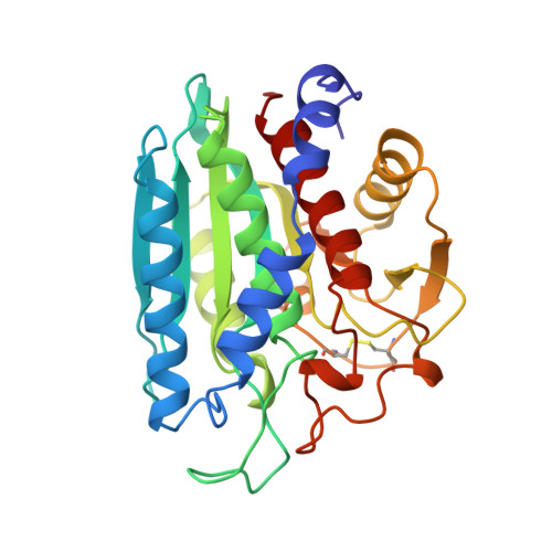
The structure of the Aeromonas proteolytica aminopeptidase complexed with a hydroxamate inhibitor. Involvement in catalysis of Glu151 and two zinc ions of the co-catalytic unit.

Chevrier, B.D'Orchymont, H.Schalk, C.Tarnus, C.Moras, D.

(1996) Eur J Biochem 237: 393-398

  • DOI: https://doi.org/10.1111/j.1432-1033.1996.0393k.x
  • Primary Citation of Related Structures:  
    1IGB

  • PubMed Abstract: 

    The structure of the complex of Aeromonas proteolytica aminopeptidase, a two-zinc exopeptidase, with the inhibitor p-iodo-D-phenylalanine hydroxamate has been determined by X-ray crystallography. Refinement of the structure, which includes 220 water molecules, using data at 0.80-0.23-nm resolution resulted in a crystallographic residual R value of 16%. The hydroxamate group adopts a planar conformation whereby the two oxygen atoms interact with the zinc ions. The N-hydroxyl group of the inhibitor is located between the two zinc ions, a position which is close to that occupied by a water molecule in the native structure. The carbonyl oxygen of the inhibitor binds to Zn1, which becomes pentacoordinated while Zn2 remains tetracoordinated, in contrast to the native protein where both zinc ions were shown to be tetracoordinated and structurally equivalent. Interactions of the carboxylate oxygens of Glu151 with the hydroxamate group play an important role in the stabilization of the complex.


  • Organizational Affiliation

    Institut de Génétique et de Biologie Moléculaire et Cellulaire, CNRS-INSERM-ULP, Illkirch, France.


Macromolecules
Find similar proteins by:  (by identity cutoff)  |  3D Structure
Entity ID: 1
MoleculeChains Sequence LengthOrganismDetailsImage
AMINOPEPTIDASE291Vibrio proteolyticusMutation(s): 0 
EC: 3.4.11.10
UniProt
Find proteins for Q01693 (Vibrio proteolyticus)
Explore Q01693 
Go to UniProtKB:  Q01693
Entity Groups  
Sequence Clusters30% Identity50% Identity70% Identity90% Identity95% Identity100% Identity
UniProt GroupQ01693
Sequence Annotations
Expand
  • Reference Sequence
Small Molecules
Binding Affinity Annotations 
IDSourceBinding Affinity
IPO Binding MOAD:  1IGB Ki: 400 (nM) from 1 assay(s)
PDBBind:  1IGB Ki: 400 (nM) from 1 assay(s)
Experimental Data & Validation

Experimental Data

  • Method: X-RAY DIFFRACTION
  • Resolution: 2.30 Å
  • R-Value Free: 0.241 
  • R-Value Work: 0.160 
  • R-Value Observed: 0.160 
  • Space Group: P 61 2 2
Unit Cell:
Length ( Å )Angle ( ˚ )
a = 109.15α = 90
b = 109.15β = 90
c = 98.35γ = 120
Software Package:
Software NamePurpose
XDSdata scaling
CCP4data reduction
X-PLORmodel building
X-PLORrefinement
XDSdata reduction
CCP4data scaling
ROTAVATA)data scaling
X-PLORphasing

Structure Validation

View Full Validation Report


Ligand Structure Quality Assessment 


Entry History 

Deposition Data

Revision History  (Full details and data files)

  • Version 1.0: 1996-08-01
    Type: Initial release
  • Version 1.1: 2008-03-24
    Changes: Version format compliance
  • Version 1.2: 2011-07-13
    Changes: Version format compliance