1IE8

Crystal Structure Of The Nuclear Receptor For Vitamin D Ligand Binding Domain Bound to KH1060


Experimental Data Snapshot

  • Method: X-RAY DIFFRACTION
  • Resolution: 1.52 Å
  • R-Value Free: 0.230 
  • R-Value Work: 0.212 

wwPDB Validation   3D Report Full Report


Ligand Structure Quality Assessment 


This is version 1.4 of the entry. See complete history


Literature

Crystal structures of the vitamin D receptor complexed to superagonist 20-epi ligands.

Tocchini-Valentini, G.Rochel, N.Wurtz, J.M.Mitschler, A.Moras, D.

(2001) Proc Natl Acad Sci U S A 98: 5491-5496

  • DOI: https://doi.org/10.1073/pnas.091018698
  • Primary Citation of Related Structures:  
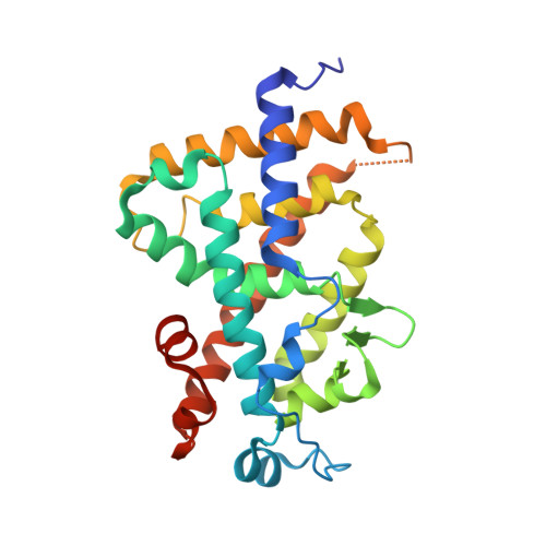
    1IE8, 1IE9

  • PubMed Abstract: 

    The crystal structures of the ligand-binding domain (LBD) of the vitamin D receptor complexed to 1alpha,25(OH)(2)D(3) and the 20-epi analogs, MC1288 and KH1060, show that the protein conformation is identical, conferring a general character to the observation first made for retinoic acid receptor (RAR) that, for a given LBD, the agonist conformation is unique, the ligands adapting to the binding pocket. In all complexes, the A- to D-ring moieties of the ligands adopt the same conformation and form identical contacts with the protein. Differences are observed only for the 17beta-aliphatic chains that adapt their conformation to anchor the 25-hydroxyl group to His-305 and His-397. The inverted geometry of the C20 methyl group induces different paths of the aliphatic chains. The ligands exhibit a low-energy conformation for MC1288 and a more strained conformation for the two others. KH1060 compensates this energy cost by additional contacts. Based on the present data, the explanation of the superagonist effect is to be found in higher stability and longer half-life of the active complex, thereby excluding different conformations of the ligand binding domain.


  • Organizational Affiliation

    Laboratoire de Biologie et Génomique Structurales, Institut de Génétique et de Biologie Moléculaire et Cellulaire, Centre National de la Recherche Scientifique/Institut National de la Santé et de la Recherche Médicale/Université Louis Pasteur, France.


Macromolecules
Find similar proteins by:  (by identity cutoff)  |  3D Structure
Entity ID: 1
MoleculeChains Sequence LengthOrganismDetailsImage
VITAMIN D3 RECEPTOR259Homo sapiensMutation(s): 0 
UniProt & NIH Common Fund Data Resources
Find proteins for P11473 (Homo sapiens)
Explore P11473 
Go to UniProtKB:  P11473
PHAROS:  P11473
GTEx:  ENSG00000111424 
Entity Groups  
Sequence Clusters30% Identity50% Identity70% Identity90% Identity95% Identity100% Identity
UniProt GroupP11473
Sequence Annotations
Expand
  • Reference Sequence
Small Molecules
Ligands 1 Unique
IDChains Name / Formula / InChI Key2D Diagram3D Interactions
KH1
Query on KH1

Download Ideal Coordinates CCD File 
B [auth A]5-(2-{1-[1-(4-ETHYL-4-HYDROXY-HEXYLOXY)-ETHYL]-7A-METHYL-OCTAHYDRO-INDEN-4-YLIDENE}-ETHYLIDENE)-4-METHYLENE-CYCLOHEXANE-1,3-DIOL
C29 H48 O4
KLZOTDOJMRMLDX-YBBVPDDNSA-N
Experimental Data & Validation

Experimental Data

  • Method: X-RAY DIFFRACTION
  • Resolution: 1.52 Å
  • R-Value Free: 0.230 
  • R-Value Work: 0.212 
  • Space Group: P 21 21 21
Unit Cell:
Length ( Å )Angle ( ˚ )
a = 44.49α = 90
b = 51.87β = 90
c = 131.39γ = 90
Software Package:
Software NamePurpose
CNSrefinement
DENZOdata reduction
CCP4data scaling
CNSphasing

Structure Validation

View Full Validation Report


Ligand Structure Quality Assessment 


Entry History 

Deposition Data

Revision History  (Full details and data files)

  • Version 1.0: 2001-05-16
    Type: Initial release
  • Version 1.1: 2008-04-27
    Changes: Version format compliance
  • Version 1.2: 2011-07-13
    Changes: Version format compliance
  • Version 1.3: 2017-08-16
    Changes: Refinement description, Source and taxonomy
  • Version 1.4: 2024-02-07
    Changes: Data collection, Database references, Derived calculations